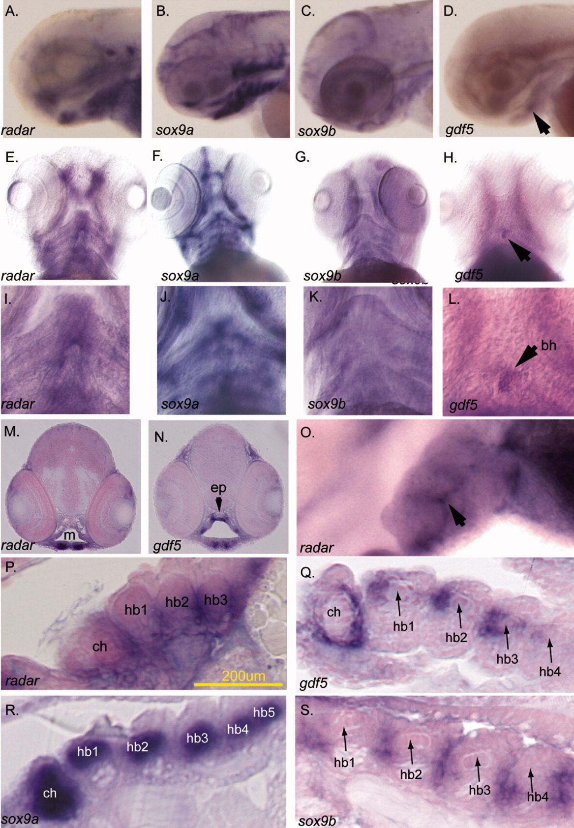
Fig. 1 AL,O: Expression of radar in the pharyngeal arch cartilages. (A-D,O lateral; E-L ventral). The expression of radar, sox9a, sox9b, and gdf5 in ventral arches is evident by in situ hybridization at 77 hours postfertilization (hpf). B,C,F,G,J,K: sox9a (B,F,J) is detected in cartilage while sox9b (C,G,K) is localized to the epithelial sheath surrounding cartilages (K). D,H,L: gdf5 is expressed in the jaw joint (white arrow, H) and medially in posterior arches, and at the basihyal (black arrows; Chiang et al.,[2001]; Yan et al.,[2005]). radar expression is detected in the jaw and along the ventral midline (E and arrows in I). M,N: Transverse sections detecting radar and gdf5 transcript. Both are expressed in the jaw joint (paired ventral staining in M and N) while only gdf5 is expressed dorsally in the pharynx (N). O: High resolution whole-mount imaging shows radar is detectable between posterior arch pharyngeal cartilages (arrow; lateral view; left = anterior). P: Sagittal section shows that radar is expressed surrounding medial hypobranchial cartilages. Q-S: Sagittal sections showing sox9a, sox9b, and gdf5 transcripts. gdf5 and radar are coexpressed near the midline around ceratohyals and hypobranchials though radar extends more ventrally below hypobranchials. bh, basihyal; ch, ceratohyal; hb1, hypobranchial 1; hb2, hypobranchial 2; hb3, hypobranchial 3; ep, ethmoid plate; e, eye; m, mouth opening.
Image
Figure Caption
Figure Data
Acknowledgments
This image is the copyrighted work of the attributed author or publisher, and
ZFIN has permission only to display this image to its users.
Additional permissions should be obtained from the applicable author or publisher of the image.
Full text @ Dev. Dyn.

