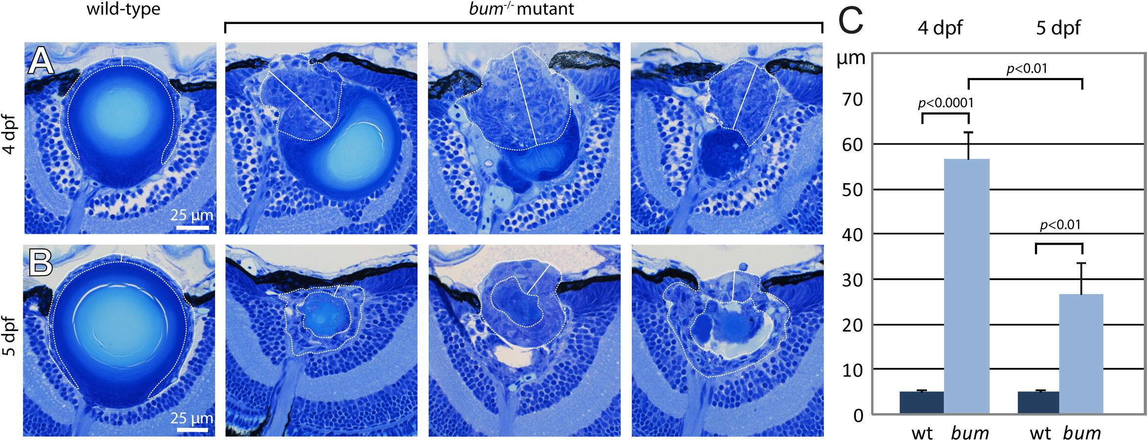
Fig. 5 The hyperproliferation of the lens epithelium regresses between 4 and 5 dpf in bum-/- mutant larvae. (A?B) Histological sections of sibling (wild-type, wt) and bum-/- mutant eyes at 4 dpf (A) and 5 dpf (B). The lens epithelial cells are encircled (curved dotted lines) and the lines along which the thickness of the lens epithelium was measured are indicated (straight solid lines). Note that the epithelium in the wild-type lenses always comprises a single layer of cells (epithelial monolayer), whereas in the mutant lenses it can be very significantly expanded (three representative examples are shown for each developmental stage). (C) Quantitative measurements of the thicknesses of wild-type (n = 10) and bum-/- mutant lens epithelia (n = 10 for 4 dpf; n = 9 for 5 dpf). Mutant epithelia are on average approx. 10-fold and 5-fold thicker than wild-type epithelia at 4 and 5 dpf, respectively. Notably, there is a statistically significant decrease of 53% in the thickness between mutant lens epithelia from 4 to 5 dpf. Error bars represent the standard error of the mean (SEM).
Reprinted from Mechanisms of Development, 127(3-4), Schonthaler, H.B., Franz-Odendaal, T.A., Hodel, C., Gehring, I., Geisler, R., Schwarz, H., Neuhauss, S.C., and Dahm, R., The zebrafish mutant bumper shows a hyperproliferation of lens epithelial cells and fibre cell degeneration leading to functional blindness, 203-219, Copyright (2010) with permission from Elsevier. Full text @ Mech. Dev.

