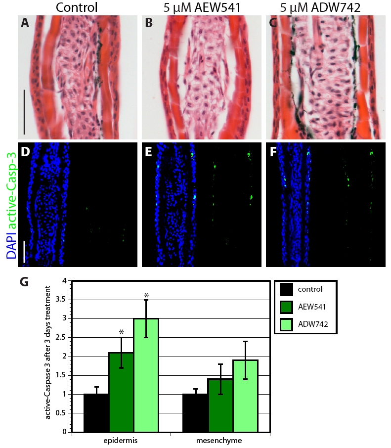
Fig. S4 Inhibition of IGF signaling increases cell apoptosis in uninjured epidermis. (A-C) Hematoxylin-Eosin stained rays of uninjured fins that were treated with 0.05% DMSO (A), NVP-AEW541 (B) or NVP-ADW742 (C) do not display differences in morphology of the epidermis. (D-F) Sections of uninjured fins stained with active-Caspase-3 antibody in green and with the nuclear marker DAPI in blue. IGF-1R inhibitor-treated fins (E,F) contain a few more active-Caspase-3-positive cells in the epidermis than in the control (D). (G) Quantification of active-Caspase-3 in uninjured fins treated with IGF-1R inhibitors for 3 days. n=6; P<0.01. Scale bars: 50 μm.
Image
Figure Caption
Acknowledgments
This image is the copyrighted work of the attributed author or publisher, and
ZFIN has permission only to display this image to its users.
Additional permissions should be obtained from the applicable author or publisher of the image.
Full text @ Development

