
|
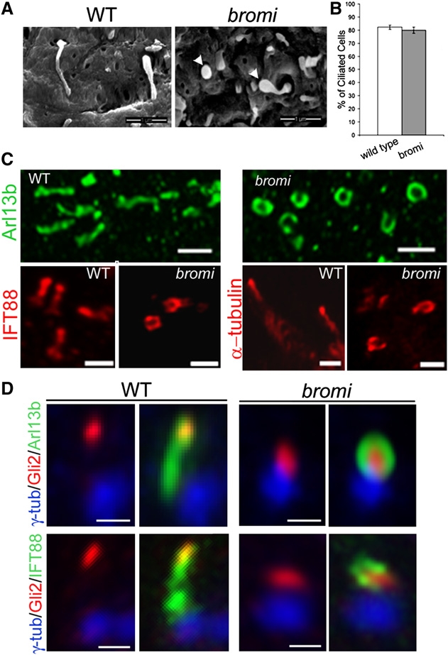
Fig. 5
Cilia Defects in bromi Mutants
(A) Neuroepithelial cilia from E10.5 WT and bromi mutant neural tubes analyzed by scanning electron microscopy. bromi mutant cilia showed a swollen or bulbous morphology (arrowheads). Scale bars, 1 μm.
(B) Cilia frequency in the WT and bromi mutant neuroepithelium. Error bars represent ± 1 SEM.
(C) Confocal images of neural tube cilia stained with Arl13b (upper panels), IFT88 and acetylated α-tubulin (lower panels). Scale bars, 1 μm.
(D) Neural tube cilia from E10.5 WT and bromi mutant embryos immunostained for γ-tubulin/Arl13b/ Gli2 or γ-tubulin/IFT88/Gli2. Scale bars, 0.5 μm.
Reprinted from Developmental Cell, 18(2), Ko, H.W., Norman, R.X., Tran, J., Fuller, K.P., Fukuda, M., and Eggenschwiler, J.T., Broad-Minded Links Cell Cycle-Related Kinase to Cilia Assembly and Hedgehog Signal Transduction, 237-247, Copyright (2010) with permission from Elsevier. Full text @ Dev. Cell