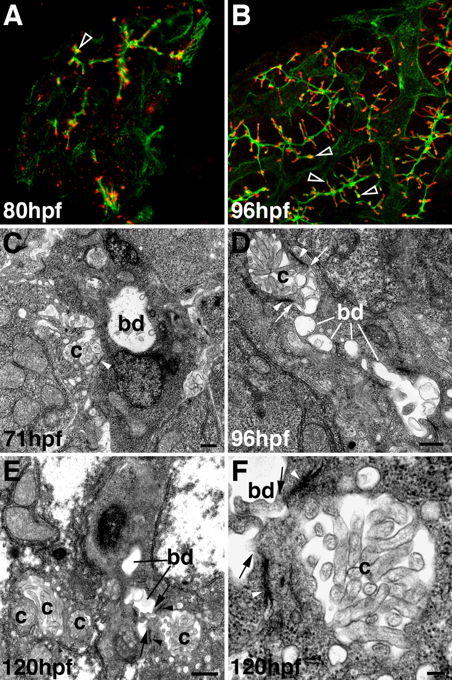
Fig. S2 Development of hepatocyte canaliculi and intrahepatic biliary network. A, B: Confocal projections through the liver of an 80-hpf (A) and 96-hpf (B) larva immunostained with Keratin-18 (green) and Mdr (red) antibodies. Compared with double immunostains of 5-dpf larvae (Fig. 2F″), there is much less overlap of the Mdr-1 and Keratin-18 epitopes at these developmental stages (open arrowheads). C-F: Transmission electron micrographs showing the canalicular-terminal duct junction (arrows) in 71-hpf (C), 96-hpf (D), and 120-hpf (E, F) larvae. bd, bile duct; c, canaliculus; arrowhead, tight junctions. Bar = 500 nm in C-E, 100 nm in F
Image
Figure Caption
Acknowledgments
This image is the copyrighted work of the attributed author or publisher, and
ZFIN has permission only to display this image to its users.
Additional permissions should be obtained from the applicable author or publisher of the image.
Full text @ Dev. Dyn.

