Image
Figure Caption
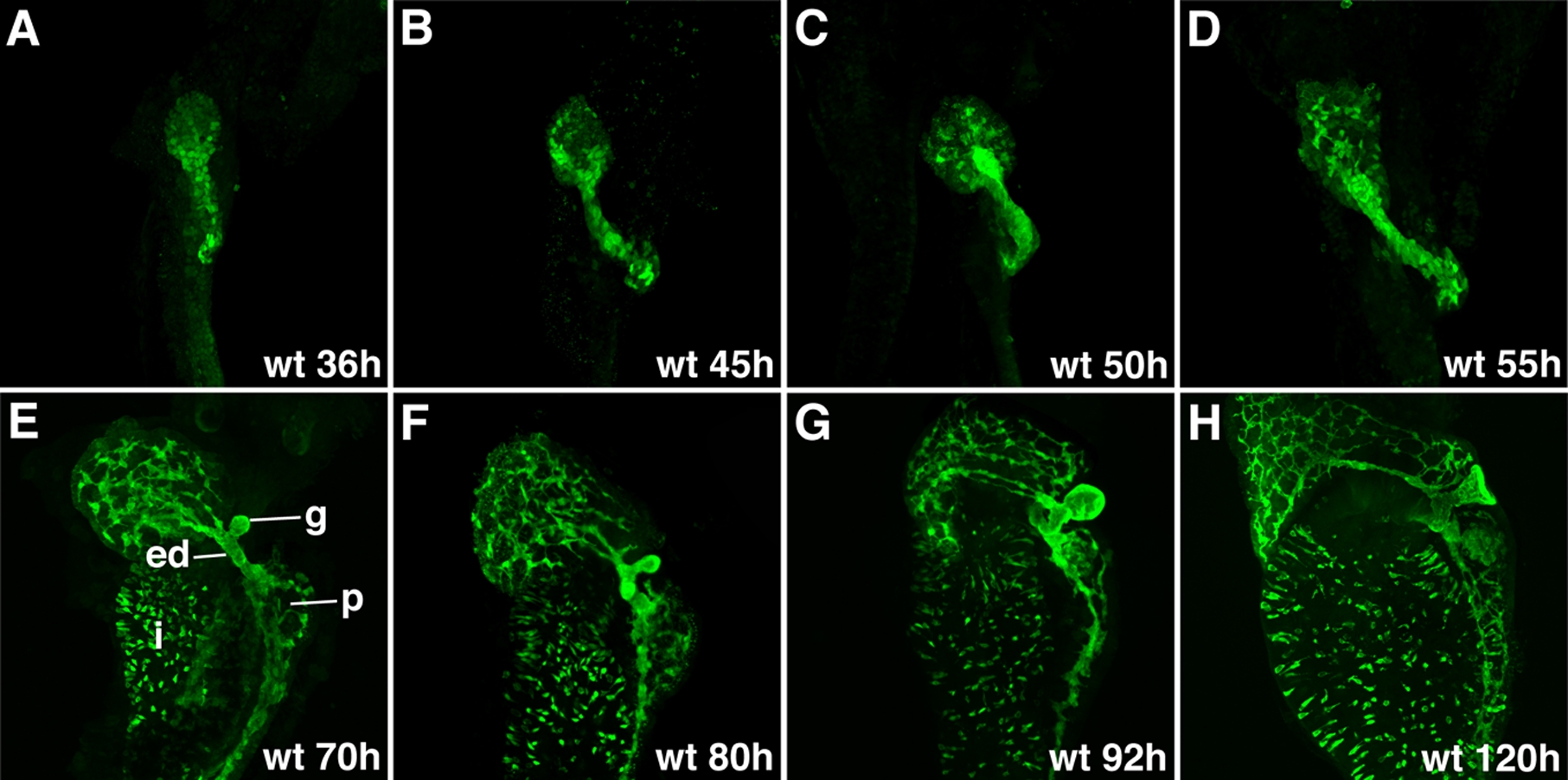
Fig. S1 Development of the intrahepatic and extrahepatic biliary system: Confocal projections through the liver of embryos and larvae immunostained with the 2F11 antibody. These images outline liver morphogenesis and development of the gallbladder (g) and extrahepatic ducts (ed) in relation to the intrahepatic ductal network. The 2F11 epitope is also detected in secretory cells on the intestinal epithelium (i), the ductal network of the pancreas (p).
Figure Data
Acknowledgments
This image is the copyrighted work of the attributed author or publisher, and
ZFIN has permission only to display this image to its users.
Additional permissions should be obtained from the applicable author or publisher of the image.
Full text @ Dev. Dyn.

