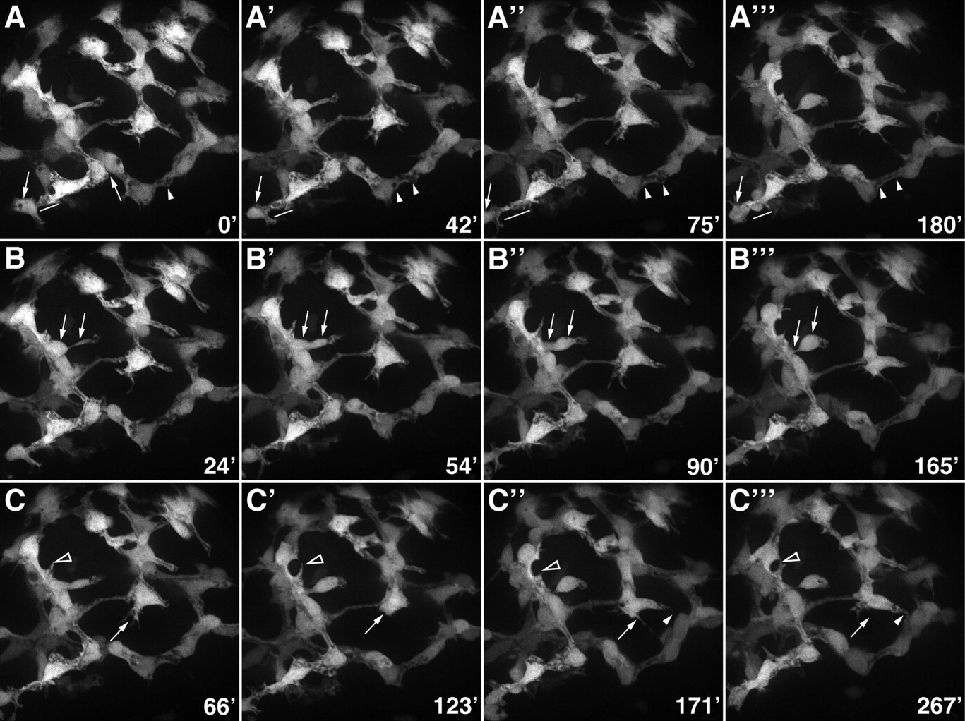
Fig. 8 Morphogenesis of the intrahepatic biliary network. Confocal projections through the liver that was dissected from a 76-hpf Notch reporter larva and cultured for 4.5 hr. Numbers in the bottom right corner indicate duration of culture in minutes. A: These images show progressive lengthening and then shortening of the cytoplasmic bridge between two adjacent biliary cells (arrows). Fusion of cytoplasmic vesicles leading to the formation of a ductal lumen within two adjacent cells is also evident (arrowheads). B: These images show the migration of a single biliary cell via a cytoplasmic extension (arrows). C: These images show the dynamic pattern of filopodia extension and retraction from two biliary cells (arrows and arrowheads).
Image
Figure Caption
Acknowledgments
This image is the copyrighted work of the attributed author or publisher, and
ZFIN has permission only to display this image to its users.
Additional permissions should be obtained from the applicable author or publisher of the image.
Full text @ Dev. Dyn.

