Image
Figure Caption
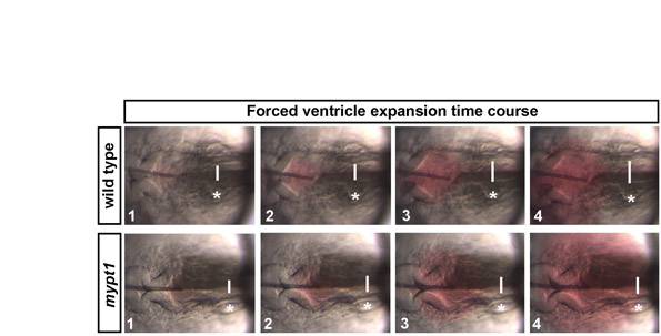
Fig. S6 The expandability of the hindbrain neuroepithelium. Time series of images in wild type or mypt1 mutants before and during forced inflation. The wild-type neuroepithelium was expanded to 1.5 times the original size by fluid injection, whereas mypt1 mutant neuroepithelium was only expanded to 1.3 times its original size. White lines indicate point where the hindbrain ventricle width was measured. Asterisks indicate the ear. Anterior is to the left in all images.
Acknowledgments
This image is the copyrighted work of the attributed author or publisher, and
ZFIN has permission only to display this image to its users.
Additional permissions should be obtained from the applicable author or publisher of the image.
Full text @ Development

