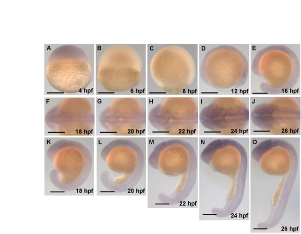
Fig. S2 mypt1 is expressed throughout the brain during hindbrain ventricle formation. In situ hybridization showing the expression of mypt1 over time, including 18-24 hpf, during which the hindbrain ventricle forms. The in situ probe was constructed from an mypt1 cDNA fragment from 132 to 3228 bp. Forward primer (from +123 to +144 relative to the start site), 5′-CGAAGGTGAAGTTCGACGATG-3′ and reverse primer (+3228 to +3248), 5′-TGTCGCCATTGTCTCGTGTT-3′. This fragment was subcloned into pGEM using the pGEM T-Easy Vector System Kit (Promega). In situ hybridization was conducted using standard procedures. (A-E) Lateral views at the stages indicated. (F-J) Dorsal views at the stages indicated. (K-O) Lateral views of the corresponding dorsal views from embryos in panels directly above. Scale bars: 200 μm.
Image
Figure Caption
Acknowledgments
This image is the copyrighted work of the attributed author or publisher, and
ZFIN has permission only to display this image to its users.
Additional permissions should be obtained from the applicable author or publisher of the image.
Full text @ Development

