
Image
|
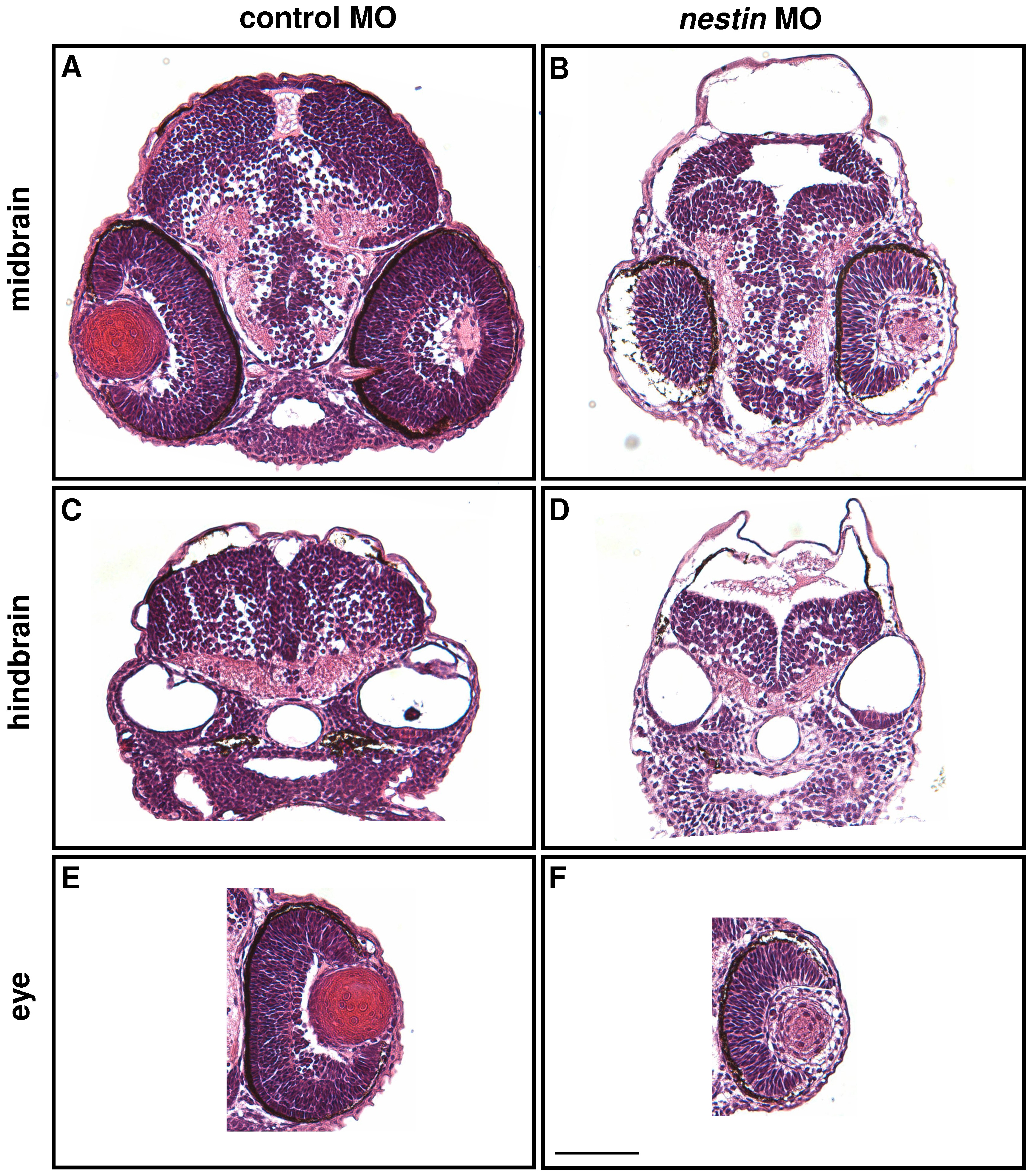
Figure Caption
Fig. 4 Examination of brain tissues and eyes by H&E staining.
Embryos at 48 hpf after control or nestin MO injection were sectioned at midbrain (A and B) and hindbrain (C and D) levels. Tissues were stained with H&E. Eyes at the midbrain level were visualized and shown in E and F. The scale bar is 50 μm.
Figure Data
Acknowledgments
This image is the copyrighted work of the attributed author or publisher, and
ZFIN has permission only to display this image to its users.
Additional permissions should be obtained from the applicable author or publisher of the image.
Full text @ PLoS One