
Image
|
Figure Caption
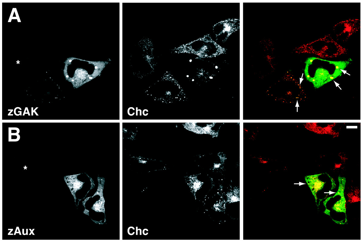
Fig. 3 The subcellular distributions of zebrafish auxilin family proteins. Spinning disc confocal micrographs of HeLa cells transfected with (A) pCS2-GFP-zGAK and (B) pCS2-GFP-zAux. The cells were also stained for clathrin heavy chain (red). The low GFP-expressing cells are indicated by asterisks. The large GFP- and clathrin-positive aggregates are indicated by arrows. Scale Bar, 10 μm.
Acknowledgments
This image is the copyrighted work of the attributed author or publisher, and
ZFIN has permission only to display this image to its users.
Additional permissions should be obtained from the applicable author or publisher of the image.
Full text @ BMC Dev. Biol.