Image
Figure Caption
Fig. S2
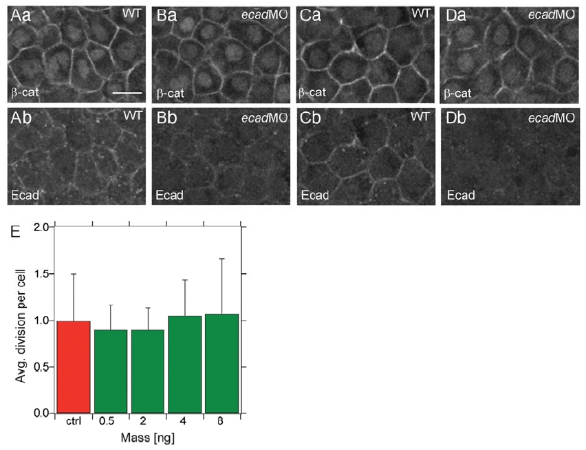
Antibody Staining Against β-Catenin and E-Cadherin in Wild-Type and e-cadherin Morphant Embryos
(Aa-Db) Epiblast (ectoderm) cells (Aa-Bb) and hypoblast (mesendoderm) cells (Ca-Db) expressing β-catenin (Aa-Da) and E-cadherin (Ab-Db) in wild-type (WT; Aa, Ab, Ca, and Cb) and e-cadherin morphant embryo (ecadMO; Ba, Bb, Da, and Db) at 7 hpf. Scale bar in (A) represents 12.5 μm.
(E) Average number of cell divisions of transplanted control and e-cadherin morphant cells (4 ng MO/embryo) during gastrulation (from 6 hpf to 10 hpf).
Acknowledgments
This image is the copyrighted work of the attributed author or publisher, and
ZFIN has permission only to display this image to its users.
Additional permissions should be obtained from the applicable author or publisher of the image.
Full text @ Curr. Biol.

