
|
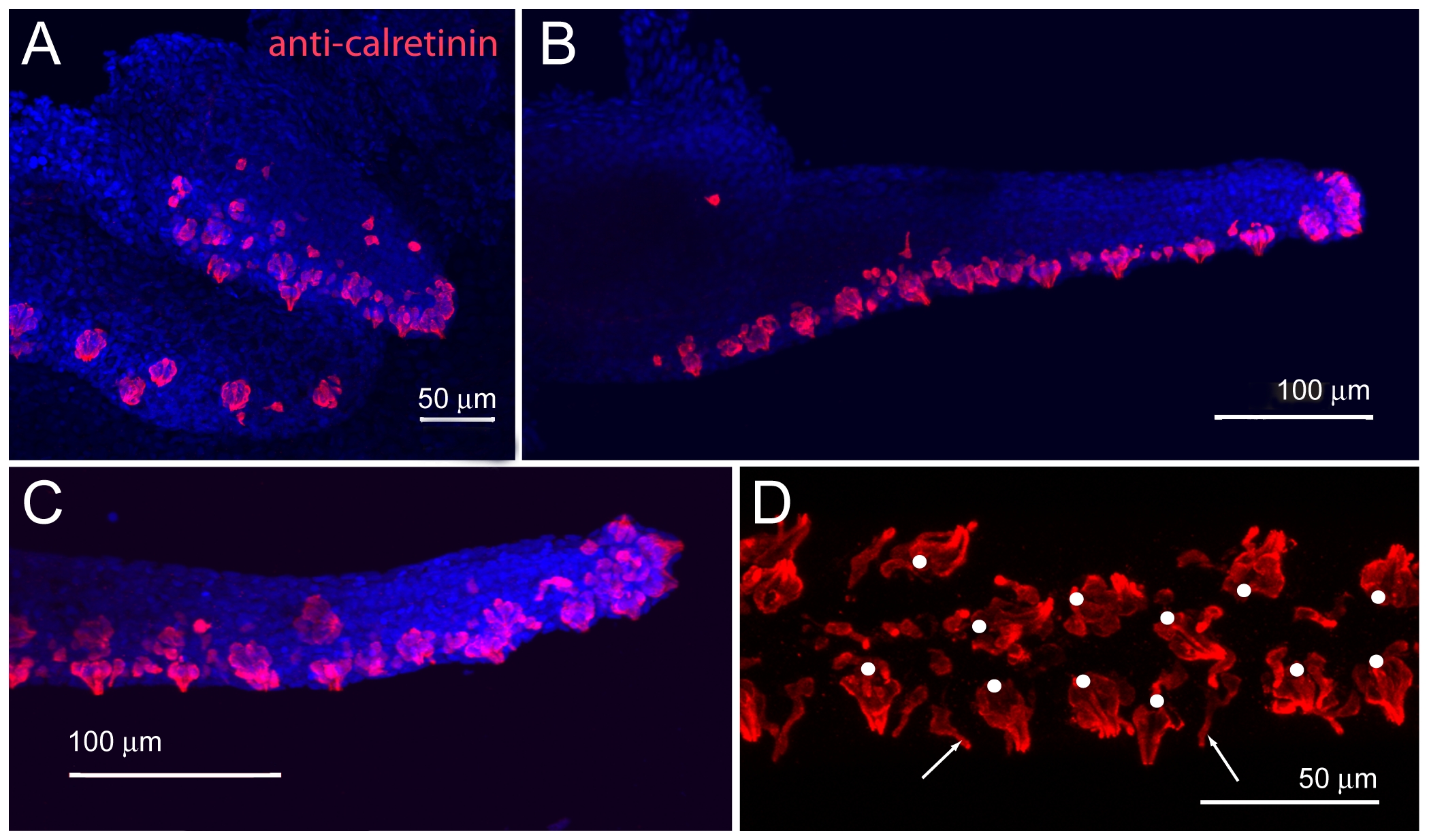
Fig. 6 Maxillary barbel taste bud development.
A) 150 μm barbel. Whole-mount immunohistochemistry (anti-calretinin) shows numerous differentiated taste buds (red) on the ventral surface and distal tip of the early barbel bud. Nuclei are counterstained blue (DAPI stain). B) 400 μm barbel. Teardrop-shaped clusters of calretinin positive cells line the ventral surface. C) Magnification of the maxillary barbel tip. D) Ventral view of the mature maxillary barbel. Teardrop-shaped taste buds (white dots) are arranged in pairs along the ventral surface. Scattered solitary chemosensory cells (SCCs, white arrows) are visible between the taste bud clusters.