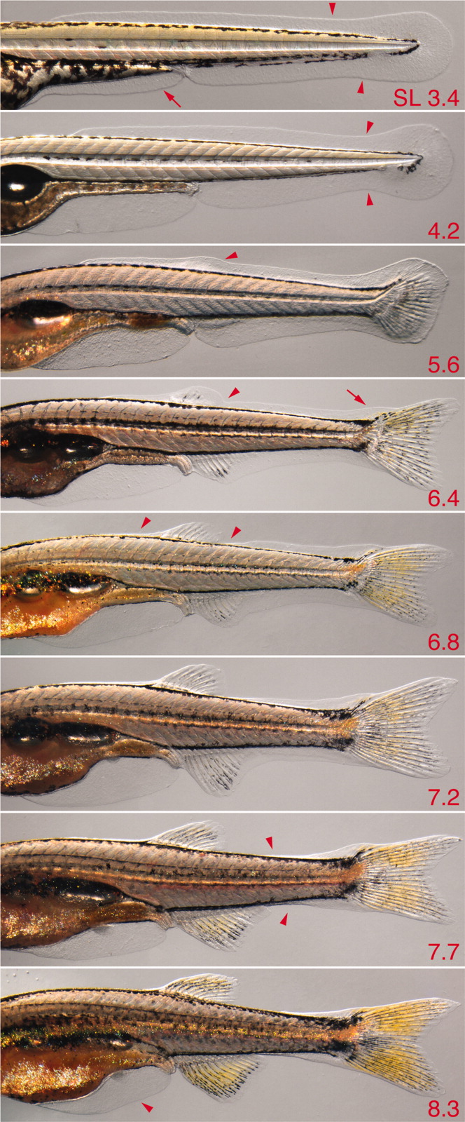
Fig. 23 Larval fin fold and fin fold resorption. Multiple individuals are shown (standard length [SL] at lower right). 3.4, Initial shapes of fin fold major lobe (arrowheads) and minor lobe (arrow); 4.2, a constriction is evident at the posterior tail (arrowhead); 5.6, a bulge is evident in the dorsal fin fold above the dorsal fin mesenchymal condensation (arrowhead); 6.4, a notch posterior to the dorsal fin indicates early fin fold resorption (arrowhead) and a bulge is evident over the developing supranotochordal fin rays (arrow); 6.8, resorption continues both anterior and posterior to the dorsal fin (arrowheads); 7.7, resorption occurs in an increasingly posterior zone along the tail (arrowheads); 8.3, early resorption of the minor lobe is revealed by flattening of its ventral posterior margin (arrowhead; also see Fig. 22).
Image
Figure Caption
Acknowledgments
This image is the copyrighted work of the attributed author or publisher, and
ZFIN has permission only to display this image to its users.
Additional permissions should be obtained from the applicable author or publisher of the image.
Full text @ Dev. Dyn.

