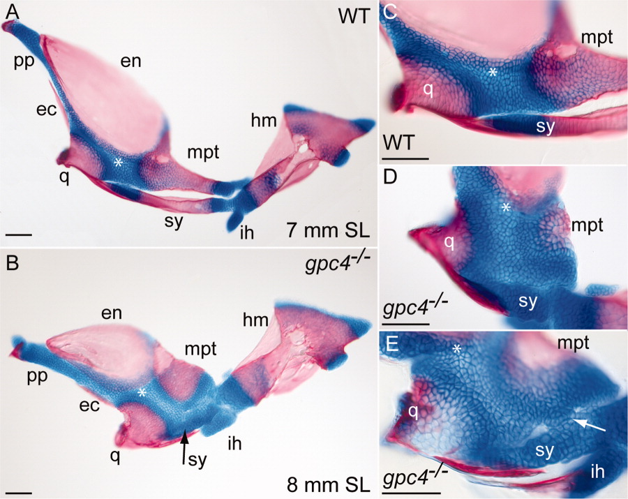
Fig. 5 Intermediate stages of symplectic bone development in gpc4-/- larvae. A: Flat-mounted facial bones and cartilages from a 7-mm wild type larva (SL = standard length). Anterior is to the left. At this stage, the symplectic (sy) is a separate bone with two cartilaginous ends. B: The corresponding region of an 8-mm gpc4-/- larva. The reduced symplectic cartilage (sy, black arrow) has not ossified and is fused to the adjacent palatoquadrate. All other ossification centers are present. C: Magnification of the wild type embryo in A. D: Magnified palatoquadrate cartilage in a second gpc4-/- larva. The reduced symplectic (sy) is a ball of cartilage broadly connected with the palatoquadrate cartilage (*). E: Symplectic region of a third gpc4-/- larva. A cluster of chondrocytes (arrow) forms a bridge between the reduced symplectic and the palatoquadrate. The quadrate is only partially mineralized. Abbreviations are as in Figures 1 and 4. ih, interhyal; pp, pterygoid process. Scale bar = 100 μm.
Image
Figure Caption
Acknowledgments
This image is the copyrighted work of the attributed author or publisher, and
ZFIN has permission only to display this image to its users.
Additional permissions should be obtained from the applicable author or publisher of the image.
Full text @ Dev. Dyn.

