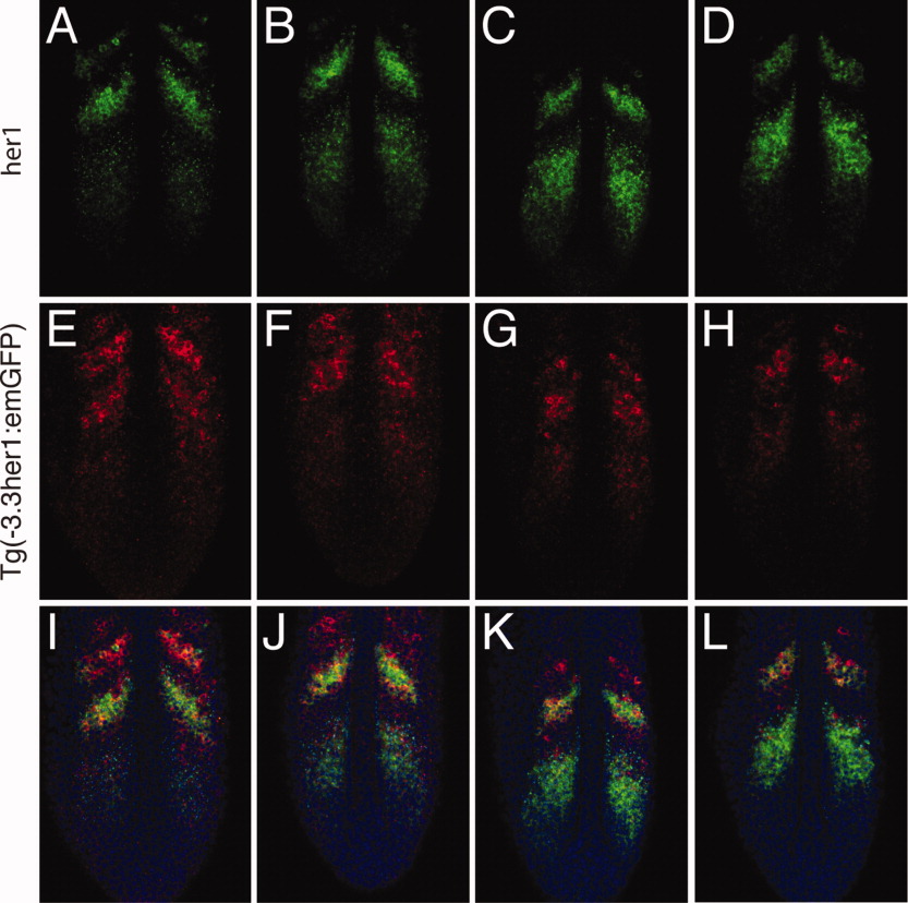
Fig. 5 The -3.3-kb enhancer does not accurately recapitulate her1 expression in the anterior presomitic mesoderm (PSM). Double fluorescent in situ hybridization of four Tg(-3.3her1:emGFP) embryos. GFP, green fluorescent protein. Three images of a single confocal section are shown for each embryo: (A,E,I), (B,F,J), (C,G,K), and (D,H,L), respectively. A-D: Expression of her1. Embryos are arranged to indicate progression of waves of her1 transcription through the somitogenesis cycle. E-H: Expression of emGFP mRNA. I-L: Merged images showing expression of her1 (green) and emGFP (red). Nuclei stained with propidium iodide are colored blue.
Image
Figure Caption
Acknowledgments
This image is the copyrighted work of the attributed author or publisher, and
ZFIN has permission only to display this image to its users.
Additional permissions should be obtained from the applicable author or publisher of the image.
Full text @ Dev. Dyn.

