
|
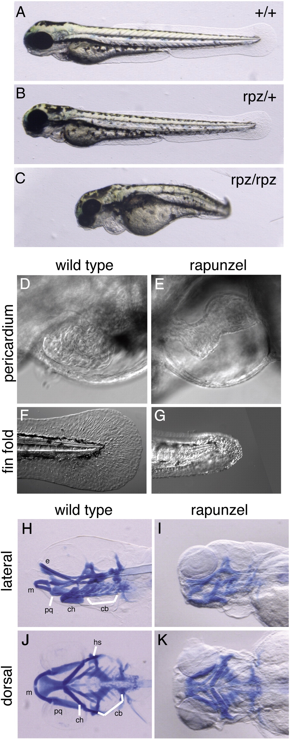
Fig. 1 Phenotype of rapunzel embryos. A–C. Wild type, heterozygous rapunzel and homozygous rapunzel embryos. D–G. Nomarski images of wild type (D, F) and homozygous rapunzel (E, G) embryos at 72 hpf demonstrating pericardial edema (E) and fin fold defects (G) in rapunzel mutants. H–K. Lateral (H, I) and ventral (J, K) views of whole-mount Alcian blue-stained wild type (H, J) and rapunzel (I, K) homozygous mutants at 96 hpf demonstrating reduced development of the craniofacial cartilages in rapunzel mutants. Craniofacial structures labeled as in Piotrowski et al., 1996; bh: baihyal, cb: ceratobranchial, ch: ceratohyal, e: ethmoid plate, hs: hyosymplectic, m: Meckel′s cartilage, and pq: palatoquadrate.
Reprinted from Developmental Biology, 334(1), Green, J., Taylor, J.J., Hindes, A., Johnson, S.L., and Goldsmith, M.I., A gain of function mutation causing skeletal overgrowth in the rapunzel mutant, 224-234, Copyright (2009) with permission from Elsevier. Full text @ Dev. Biol.