
|
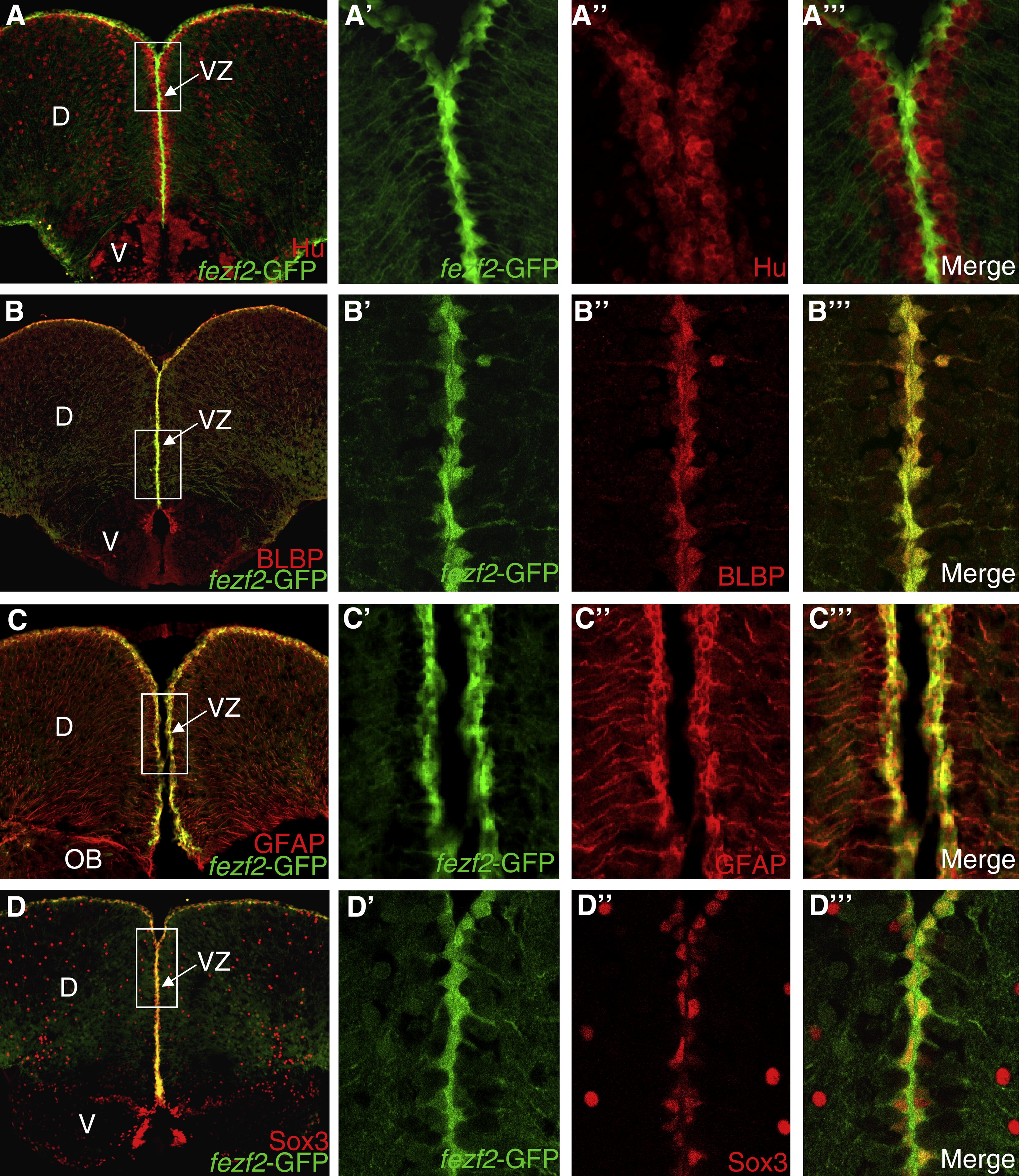
Fig. 3a fezf2 is expressed in radial glial cells of the telencephalic ventricular zone, which colocalize with markers of neural stem cells and proliferation. (A) Coronal section through telencephalon showing double-label of fezf2-GFP (green) and Hu (red) (20x magnification). (A′?A′″) Closer view of the boxed region (100x magnification) shows that fezf2-GFP+ cells have radial glial morphology (A′) and do not overlap with neuronal marker HuC/D (A″′). (B) Coronal section through telencephalon showing colocalization of fezf2-GFP (green) and BLBP (red) in the pallial ventricular zone (20x magnification). (B′?B″′) Single confocal Z-section (∼0.5 μm) of the boxed region (100x magnification) shows that fezf2-GFP expression colocalizes precisely with BLBP+ radial glial cells. (C) Coronal section through anterior telencephalon showing double-label of fezf2-GFP (green) and GFAP (red) (20x magnification). (C′?C′″) Single confocal Z-section of the boxed region (40x magnification) shows that fezf2-GFP+ cells colocalize with neural stem cell marker GFAP. (D) Coronal section through telencephalon showing colocalization of fezf2-GFP (green) and Sox3 (red) in the pallial ventricular zone (20x magnification). (D′?D″′) Single confocal Z-section of the boxed region shows that fezf2-GFP+ radial glial cells colocalize with neural stem cell marker Sox3 (100x magnification).
Reprinted from Gene expression patterns : GEP, 9(6), Berberoglu, M.A., Dong, Z., Mueller, T., and Guo, S., fezf2 expression delineates cells with proliferative potential and expressing markers of neural stem cells in the adult zebrafish brain, 411-422, Copyright (2009) with permission from Elsevier. Full text @ Gene Expr. Patterns