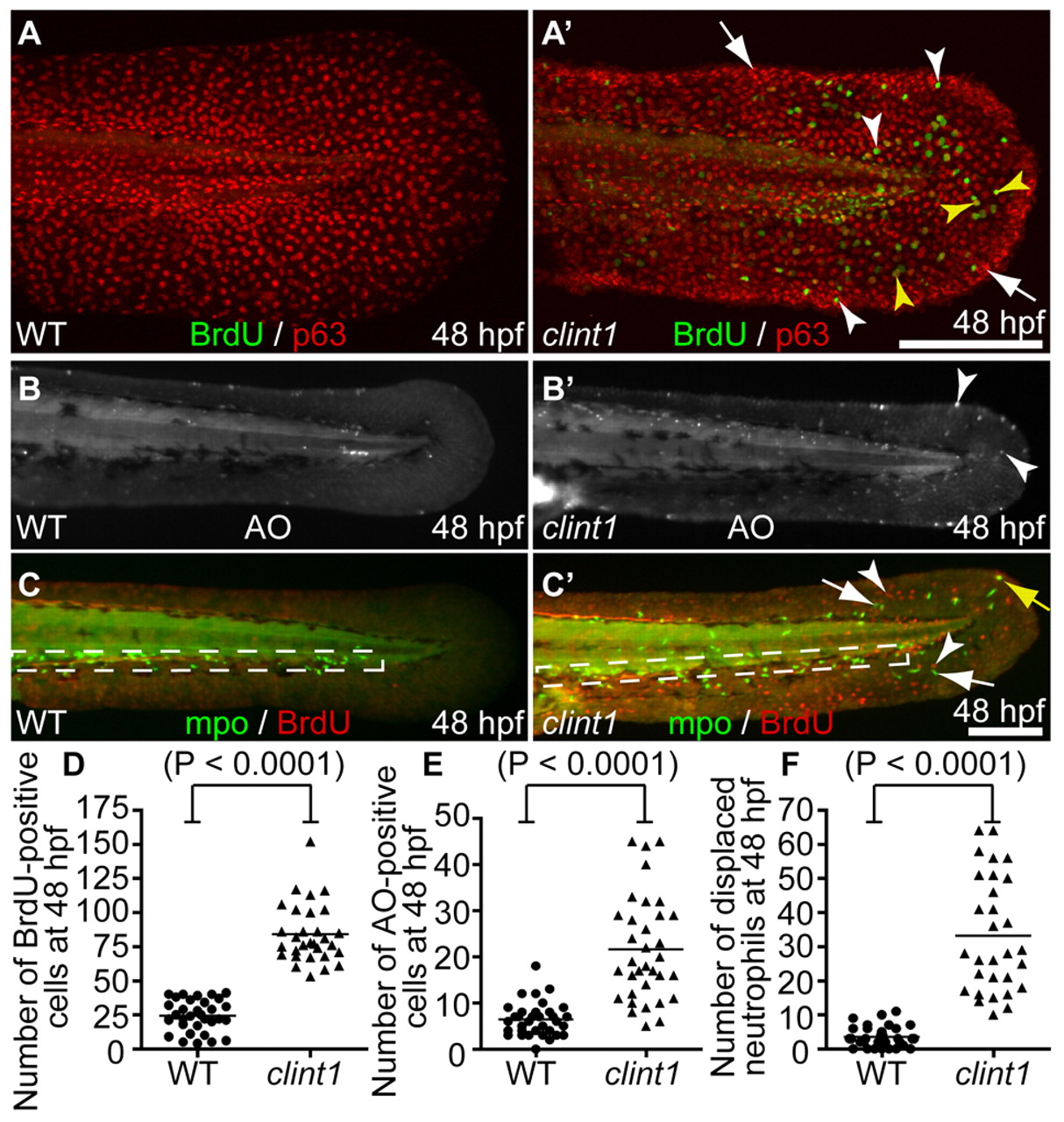
Fig. 2 hi1520 mutant phenotypes at 48 hpf. (A-C′) Wild-type (A-C) and clint1 mutant (A′-C′) 48 hpf zebrafish embryos labeled for BrdU incorporation (green) and p63 (red) (A,A′), Acridine Orange (AO) (B,B′) or Mpo (green) and BrdU incorporation (red) (C,C′). Boxed region, caudal hematopoietic tissue (CHT). Arrowheads identify proliferation in p63-positive (yellow arrowheads) and p63-negative (white arrowheads) cells (A′,C′), and cell death (B′) in clint1 mutants. Arrows identify epidermal aggregates (A′) and neutrophils (C′) in clint1 mutants. Yellow arrow (C′) identifies a proliferating neutrophil. (D-F) Quantification of BrdU (D) and AO (E) in fin fold and overall displacement of neutrophils (F) in wild-type (circles) and clint1 mutant (triangles) embryos. Bars represent the mean. Scale bars: 200 μm.
Image
Figure Caption
Figure Data
Acknowledgments
This image is the copyrighted work of the attributed author or publisher, and
ZFIN has permission only to display this image to its users.
Additional permissions should be obtained from the applicable author or publisher of the image.
Full text @ Development

