
Image
|
Figure Caption
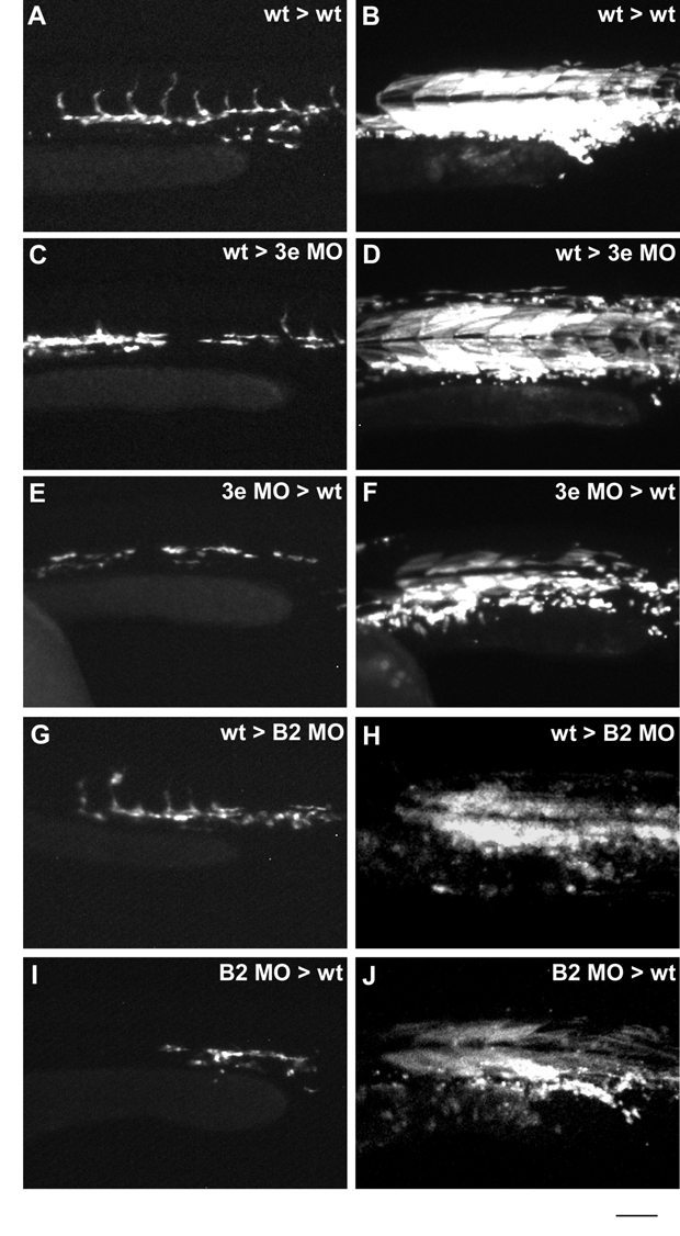
Fig. S3
Rhodamine dextran images of representative transplanted embryos from Figure 5.
(A, C, E, G, I) Tg(kdr:GFP)la116 donor angioblasts in non-transgenic recipient embryos from Fig. 5. (B, D, F, H, J) Corresponding rhodamine dextran images for embryos in A, C, E, G, I showing most transplanted cells residing in tissues of mesodermal origin. Scale bar is 100 μm.
Acknowledgments
This image is the copyrighted work of the attributed author or publisher, and
ZFIN has permission only to display this image to its users.
Additional permissions should be obtained from the applicable author or publisher of the image.
Reprinted from Developmental Biology, 331(2), Lamont, R.E., Lamont, E.J., and Childs, S.J., Antagonistic interactions among Plexins regulate the timing of intersegmental vessel formation, 199-209, Copyright (2009) with permission from Elsevier. Full text @ Dev. Biol.