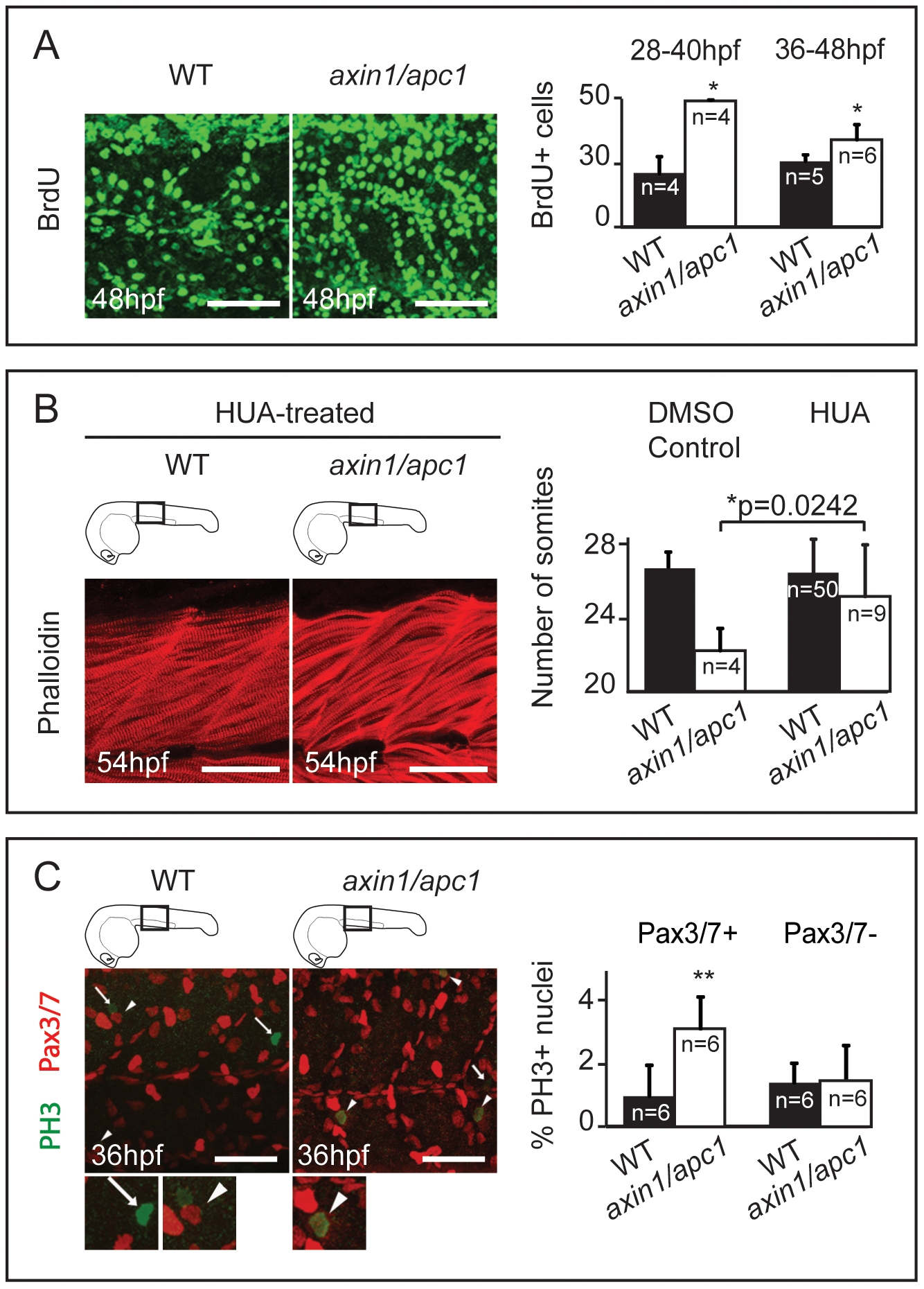
Fig. 4 Myotome hyperproliferation and sustained differentiation in axin/apc1 embryos.
(A) BrdU pulse was performed at 36 hpf, chased for 12 hours, and imaged at 48 hpf. Embryos were imaged at the level of the yolk extension. Scale bar, 25 μm. BrdU+ pulse was performed at 28 hpf or 36 hpf, and quantification of number of BrdU+ proliferating cells per somite was done 12 hours later at 40 hpf or 48 hpf, respectively. (C) HUA treatment of embryos from 24 hpf until fixation at 54 hpf. Inhibition of proliferation with HUA from 24 hpf results in rescue of muscle hypertrophy. Muscle fibers were stained with Phalloidin, and imaged at 54 hpf at the level of the yolk extension. Compare with untreated wild-types in Fig. 2A (right panels). Scale bar, 25 μm. Quantification of number of somites is increased in axin1/apc1 mutants upon HUA treatment. (E) Colocalization of Pax3/7+ and PH 3+ cells shows proliferating muscle progenitors. Quantification of proliferating Pax3/7+ and Pax3/7- cells in the wild-types vs. axin1/apc1 mutant embryos shows significantly more proliferating Pax3/7+ cells in the mutants. Scale bar, 50 μm.

