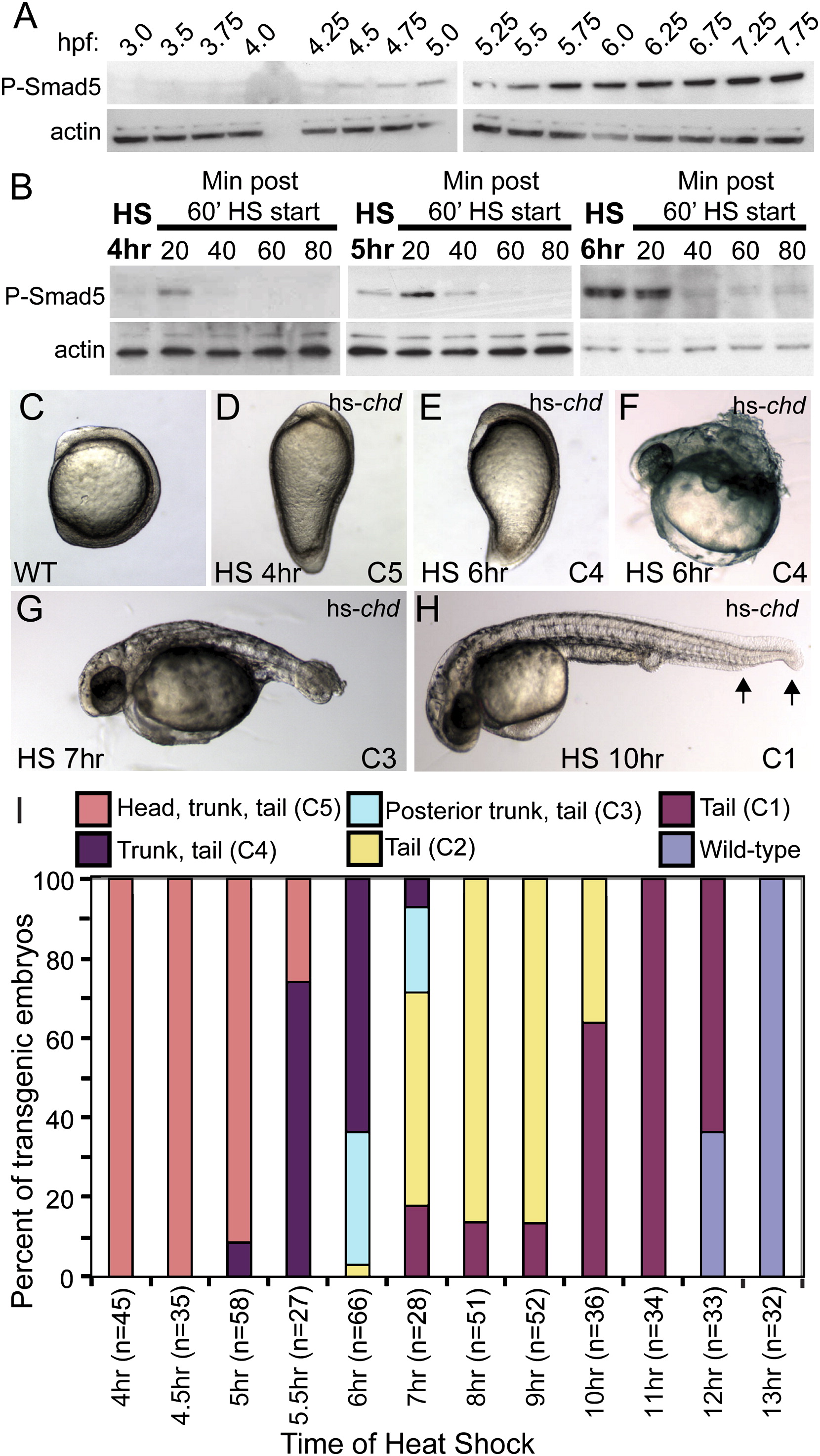
Fig. 3 Tg(hsp70:chd) Rapidly Inhibits BMP Signaling, Generating a Range of Dorsalized Phenotypes Dependent on Induction Time
(A) P-Smad5 western blot at blastula (3.0?5.25 hpf), onset of gastrulation (5.5 hpf) through midgastrulation (5.75?7.75 hpf) stages.
(B) P-Smad5 during (20 and 40 min time points) and immediately after a 60 min HS at 4, 5, or 6 hpf. Actin is a loading control.
(C?F) Nontransgenic WT (C) and transgenic siblings heat-shocked at 4 hpf (D) or 6 hpf (E) at the three-somite stage and at 1 day postfertilization (dpf) (F).
(G and H) Dorsalization at 1 dpf of embryos heat-shocked at 7 hpf (G) and 10 hpf ([H], arrows indicate loss of ventral tail tissue).
(I) Distribution of dorsalized phenotypes following HS at different time points.
Reprinted from Developmental Cell, 14(1), Tucker, J.A., Mintzer, K.A., and Mullins, M.C., The BMP signaling gradient patterns dorsoventral tissues in a temporally progressive manner along the anteroposterior axis, 108-119, Copyright (2008) with permission from Elsevier. Full text @ Dev. Cell

