
Image
|
Figure Caption
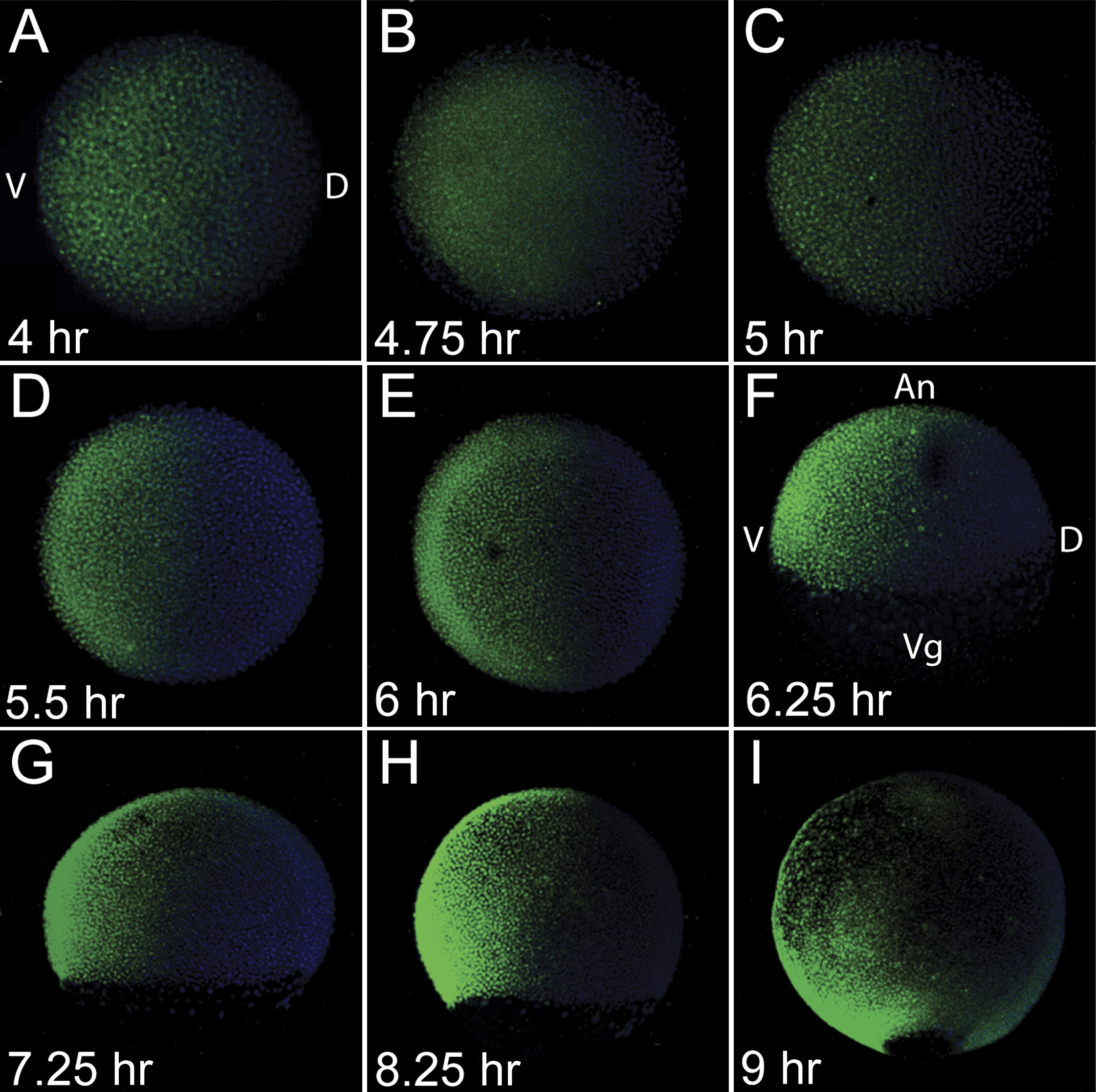
Fig. 1 Formation of the BMP Activity Gradient
Whole-mount P-Smad5 (green) antibody staining during blastula stages, at 4 hpf (A), 4.75 hpf (B), and 5 hpf (C) and during gastrula stages at 5.5 hpf (D), 6 hpf (E), 6.25 hpf (F), 7.25 hpf (G), 8.25 hpf (H), and 9 hpf (I). Nuclei were stained with DAPI (blue). (A)?(E) Animal view. (F)?(I) Lateral view. Dorsal to right in all.
Acknowledgments
This image is the copyrighted work of the attributed author or publisher, and
ZFIN has permission only to display this image to its users.
Additional permissions should be obtained from the applicable author or publisher of the image.
Reprinted from Developmental Cell, 14(1), Tucker, J.A., Mintzer, K.A., and Mullins, M.C., The BMP signaling gradient patterns dorsoventral tissues in a temporally progressive manner along the anteroposterior axis, 108-119, Copyright (2008) with permission from Elsevier. Full text @ Dev. Cell