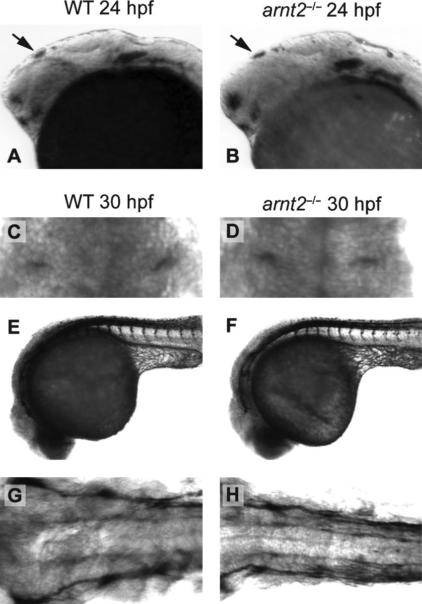
Fig. 4 Sensory neurons involved in the touch and startle responses appear normal in arnt2-/- embryos at 24–30 hpf. Lateral views for whole-mount ISH of islet1 in a representative WT larva and arnt2-/- larva is shown at 24 hpf (A, B). The pattern of islet1 staining in the brain (black arrow) is similar for the two genotypes, suggesting that regions where primary neurons form are developing normally. 3A10 IHC staining of Mauthner neurons is similar in a representative WT embryo and arnt2-/- mutant embryo at 30 hpf (C, D). ZNP1 IHC to identify primary motor neurons and trigeminal ganglia is shown in a lateral view of a WT embryo and an arnt2-/- embryo (E, F) and in a dorsal view (G, H) at 30 hpf. ZNP1 IHC is similar in the two genotypes. Results in all panels are representative of 5–9 embryos/genotype.
Image
Figure Caption
Figure Data
Acknowledgments
This image is the copyrighted work of the attributed author or publisher, and
ZFIN has permission only to display this image to its users.
Additional permissions should be obtained from the applicable author or publisher of the image.
Full text @ Zebrafish

