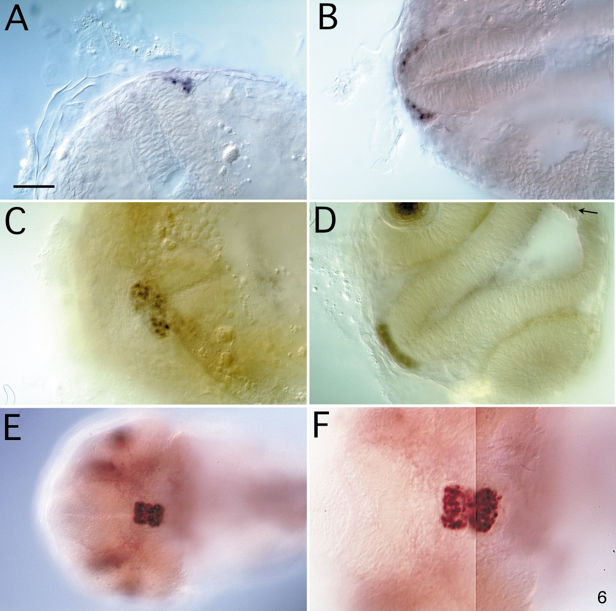
Fig. 6 Expression of Lim3 in the pituitary anlage. Embryos were stained in whole mount, dissected from the yolk, and photographed from a ventral view. The embryos are oriented so that anterior faces left, except in A where anterior faces up. The plane of focus cuts through part of the ventral diencephalon. A–D, wild-type embryos; E and F, flh mutant embryos. (A) At the 21-somite stage, Lim3 is present in approximately 8 cells, laterally on the left side of the anteriormost end of the neural tube. (B) At the 24-somite stage, cells on both sides of the neural tube express Lim3 still in a lateral position. (C) By 28 h, Lim3-expressing cells have moved to the midline and coalesced into the pituitary cluster. (D) An anterior view of a 28-h embryo. The optical section runs from the anterior edge of the pituitary cluster through the epiphysis, with the diencephalon in the plane of focus. A stained cell can be seen slightly below the focal plane in the epiphysis (arrow). The Lim3-positive cells of the pituitary cluster are one cell thick at the anterior edge. The staining in the eye lens is not nuclear and is probably artifactual. (E,F) The same flh embryo shown at two magnifications; since the enlarged pituitary was not in one plane of focus at the higher magnification, F was assembled from two images of the same embryo (see E). When comparing the wild-type embryo in C with the mutant in F, it is apparent that the flh pituitary is twice as large in the A/P dimension, but slightly more compact in the L/R dimension; there are approximately twice as many Lim3-positive cells in the mutant than in the wild-type pituitary. Scale bar is 50 μm except in E where it is 100 μm.
Reprinted from Developmental Biology, 192, Glasgow, E., Karavanov, A.A., and Dawid, I.B., Neuronal and neuroendocrine expression of lim3, a LIM class homeobox gene, is altered in mutant zebrafish with axial signaling defects, 405-419, Copyright (1997) with permission from Elsevier. Full text @ Dev. Biol.

