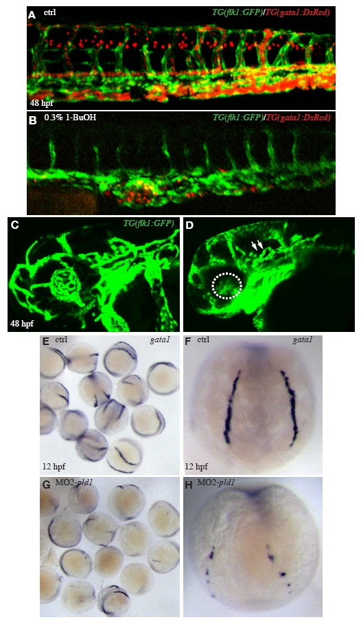
Fig. S6 Loss of Pld1 function disrupts the cranial vessels and blood cells formation. (A) At 48 hpf, Control TG(flk1:GFP)/(gata1:DsRed) embryos show a normal formation of ISV and axis vessels (green) in the trunk region (lateral view) with flowing blood cells in red, and 0.3% 1-BuOH treated embryos (B) show defective ISV formation and slightly reduced axis formation at the similar area, however blood cells are much more strongly affected. At 48 hpf, compared to control embryos (C), major cranial vessels in the head region are mainly formed in MO2-pld1 injected embryos (D), with minor disorganization of CCtA (Cerebellar central artery) (white arrows) and reduction of IOC vessels (Inner optic circle, white dotted circle) in the retinal area. At 12 hpf (6-somite stage), uninjected control embryos (E, group; F, dorsal view) express gata1 bilaterally in the lateral plate mesoderm, however the expression are strongly reduced or absent in MO2-pld1 injected embryos (G, group; H, dorsal view).
Reprinted from Developmental Biology, 328(2), Zeng, X.X., Zheng, X., Xiang, Y., Cho, H.P., Jessen, J.R., Zhong, T.P., Solnica-Krezel, L., and Brown, H.A., Phospholipase D1 is required for angiogenesis of intersegmental blood vessels in zebrafish, 363-376, Copyright (2009) with permission from Elsevier. Full text @ Dev. Biol.

