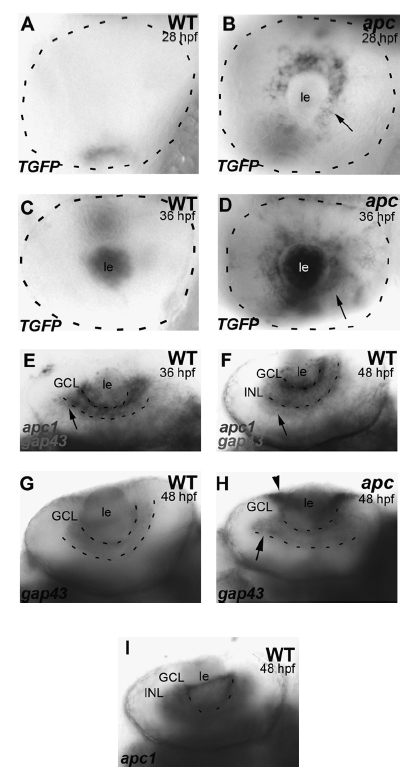
Fig. 1 The apcCA50a/CA50a mutation results in aberrant LEF/β-catenin transcription. (A, B) In wild-type embryos at 28 hpf (A), TGFP is absent from the retina. In apc mutants (B), TGFP is expressed in the retinal epithelium surrounding the lens (arrow). (C, D) At 36 hpf, TGFP is upregulated in the mutant lens and RGC layer (arrow in D). (E, F) Double in situ hybridization to apc1 and gap43, which marks differentiating neurons developing axons at 36 (E) and 48 hpf (F), shows that apc1 is expressed in areas of the retina that undergo differentiation. Dashed line indicates eye circumference (A? D) Lateral view, anterior to the left. (E, F) Ventral view, anterior to the left. (G, H) gap43 Expression in WT (G) and apc mutants (H) at 48 hpf shows disorganization of the GCL in the mutants. (I) apc1 Expression in the GCL and INL of the WT retina at 48 hpf. GCL, ganglion cell layer; INL, inner nuclear layer; le, lens; RGC, retinal ganglion cell. Scale bar = 50 μm.
Image
Figure Caption
Figure Data
Acknowledgments
This image is the copyrighted work of the attributed author or publisher, and
ZFIN has permission only to display this image to its users.
Additional permissions should be obtained from the applicable author or publisher of the image.
Full text @ Zebrafish

