
|
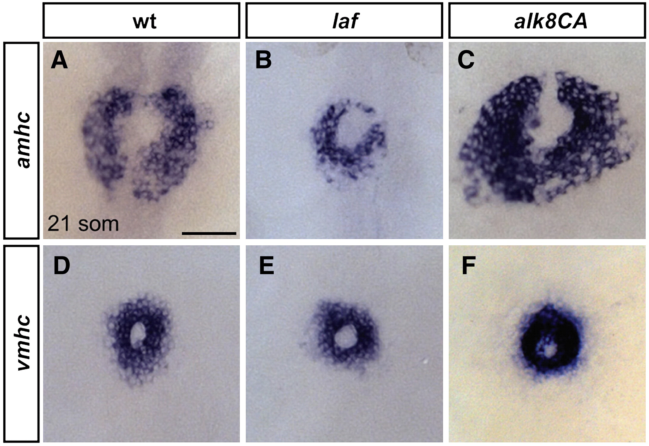
Fig. 2 Alk8 function impacts atrial cell number prior to heart tube formation. (A?F) In situ hybridization depicts expression of amhc (A?C) and vmhc (D?F) at the 21-somite stage; dorsal views, anterior to the top. Scale bar represents 50 μm. (A) In wild-type embryos, amhc is expressed in a ring of atrial cardiomyocytes just prior to heart tube extension, whereas in laf mutant embryos (B), the population of amhc-expressing cells is clearly reduced (n = 14/15). (C) In alk8CA mRNA-injected embryos displaying a v2?v3 ventralized body morphology, the population of amhc-expressing cells is clearly increased (n = 20/23). (D) vmhc is expressed in a ring of ventricular cardiomyocytes, located interior to the ring of atrial cardiomyocytes. The population of vmhc-expressing cells is comparable to wild-type in (E) laf mutants (n = 14/14) and seems slightly increased in (F) alk8CA mRNA-injected embryos (n = 22/22).
Reprinted from Developmental Biology, 328(2), Marques, S.R., and Yelon, D., Differential requirement for BMP signaling in atrial and ventricular lineages establishes cardiac chamber proportionality, 472-482, Copyright (2009) with permission from Elsevier. Full text @ Dev. Biol.