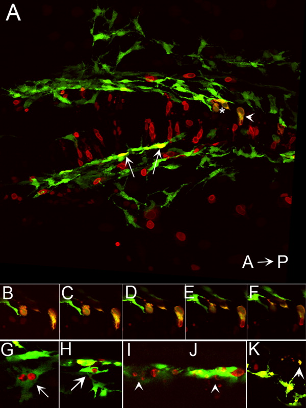
Fig. 8 Ectopic expression of foxd3 promotes cell death in kit mutants. A: Confocal stack image showing a dorsal view of the posterior head of a 28 hours postfertilization (hpf) kitw34/w34 embryo transgenic for mi:gfp and also expressing myc-foxd3 (red) under the control of a heat shock promoter. Expression of myc-foxd3 was induced at 19 hpf. Both wild-type (arrows) and fragmenting (asterisk and arrowhead) melanophores are observed. B-F: Confocal slices showing fragmenting cells indicated by asterisk and arrowhead in A. Note the presence of Foxd3-positive fragments within the GFP+ melanophores. G,H: Confocal slices showing additional examples of GFP/Foxd3+ cells designated as normal melanophores. I-K: Confocal slices showing examples of GFP/Foxd3+ cells designated as fragmenting melanophores (arrowheads).
Image
Figure Caption
Acknowledgments
This image is the copyrighted work of the attributed author or publisher, and
ZFIN has permission only to display this image to its users.
Additional permissions should be obtained from the applicable author or publisher of the image.
Full text @ Dev. Dyn.

