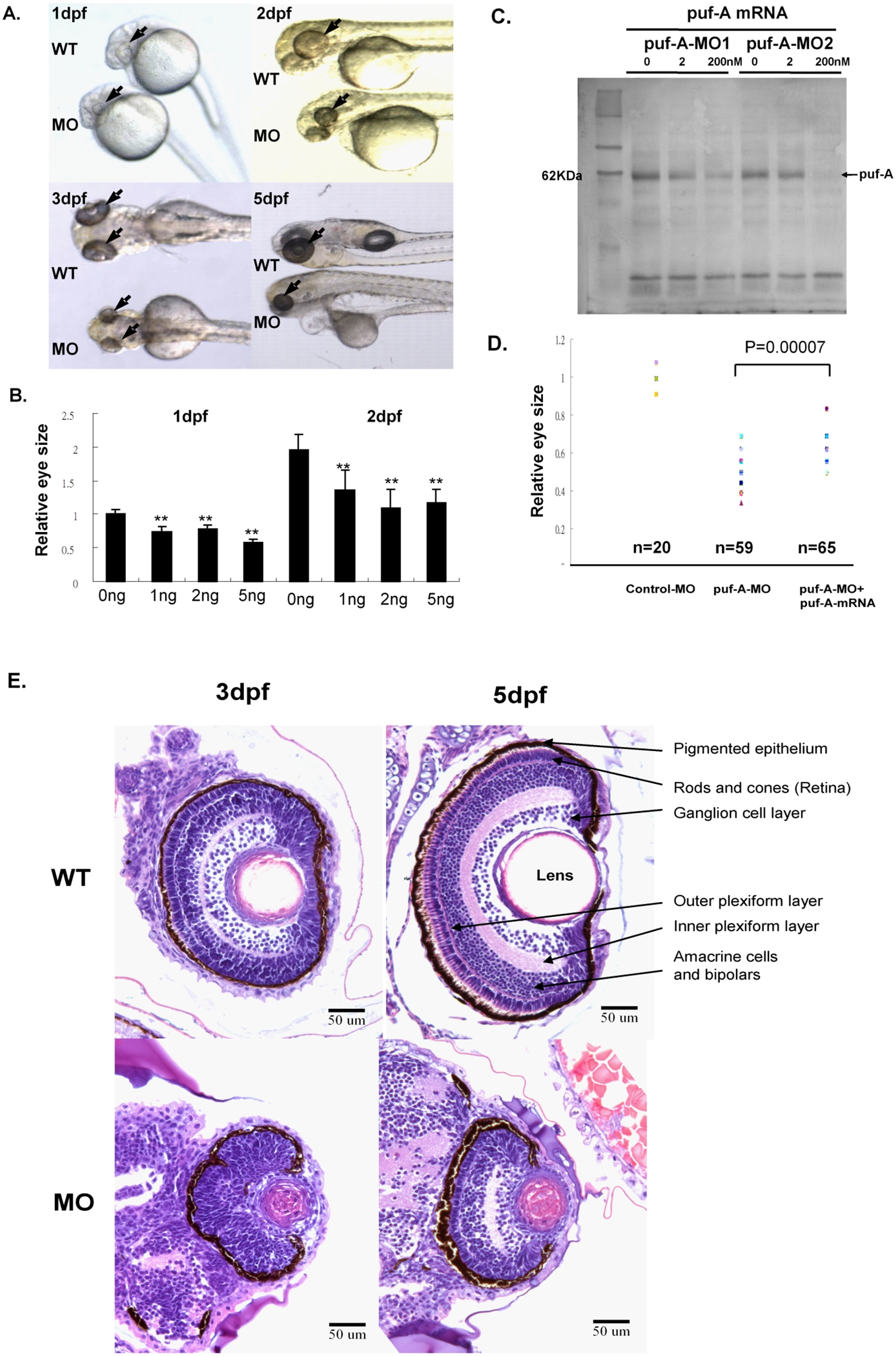
Fig. 3 The phenotypes of puf-A morphants in the zebrafish.
(A) Zebrafish embryos at the 1∼4-cell stage were treated with 5 ng puf-A morpholino (MO1) by microinjection. The phenotypes of the wild-type and morphants are shown in lateral view at 1, 2, 3, and 5 days post-fertilization (dpf) after treatment. Black arrows point to the eyes. (B) Various amounts of MO1 were microinjected into zebrafish embryos, and the eye size was measured at 1 or 2 dpf and compared to the eye size of control fish. The ?relative eye size? was defined by the value of eye size in MOs relative to the average size of eyes in normal embryos of WT fish. The average value of eye size in normal embryos at 1dpf was considered as 1. Error bars represent the standard error of the mean. ** refer to p<0.01 Student's t-test). (C) 0, 2, 200 nM puf-A-MO1 or puf-A-MO2 were added to the puf-A generated through in vitro transcription/translation reactions. One microliter of the reaction mixture was separated on 10% SDS/PAGE, blotted, incubated with streptavidin-AP, and developed with NBT-BCIP reagents. (D) The 5 ng control- or puf-A-MO1 was used for microinjection. In addition, 200 pg of capped puf-A RNA was co-injected with puf-A-MO1 to check the specificity of MO knockdown. The ?relative eye size? was defined as above. The eye size was measured at 2 dpf. Control-MO, n = 20 embryos; puf-A-MO1, n = 59; puf-A-MO1+ capped RNA, n = 65. The p value in Student's t-test for the difference between puf-A-MO and puf-A-MO+mRNA was <0.00007. (E) Transverse histological sections of zebrafish wild-type and morphant (puf-A-MO1) eyes stained with hematoxylin and eosin at 3 and 5 dpf.

