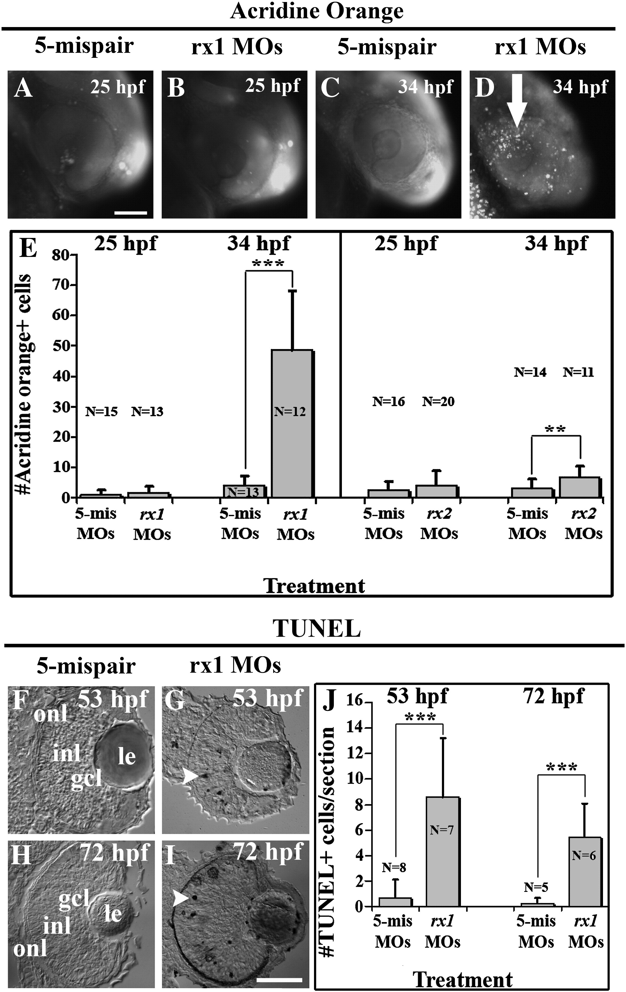
Fig. 6 Rx1 and rx2 morphants exhibit significant increases in retinal cell death. (A–D) Grayscale images of live control (A, C) and rx1 morphant (B, D) embryos stained with acridine orange showing very little cell death at 25 hpf (A, B) but a dramatic increase in cell death (C vs. D) at 34 hpf in morphants as compared to controls; arrow in D indicates acridine orange-positive cells in the eye of a morphant. E. Average numbers of acridine orange-positive cells as a function of treatment and time of assessment. Significant differences were evident following rx1 and rx2 depletion when assessed at 34 hpf (rx1,***p < 0.001; rx2,**p < 0.01) but not at 25 hpf. (F–I) Grayscale images of sectioned control (F, H) and rx1 morphant (G, I) embryos processed for the TUNEL cell death assay at 53 hpf (F, G) and 72 hpf (H, I), showing increased cell death in rx1-depleted retinas; arrowheads in G and I indicate TUNEL-positive cells. (J) Average numbers of TUNEL-positive cells as a function of treatment and time of assessment. Significant differences were evident following rx1 depletion when assessed at 53 hpf and 72 hpf (***p < 0.001). le = lens; gcl = ganglion cell layer; inl = inner nuclear layer; onl = outer nuclear layer; scale bar in A (applies to A–D) and I (applies to F–I) = 50 μm; error bars indicate ± 1 SD.
Reprinted from Developmental Biology, 328(1), Nelson, S.M., Park, L., and Stenkamp, D.L., Retinal homeobox 1 is required for retinal neurogenesis and photoreceptor differentiation in embryonic zebrafish, 24-39, Copyright (2009) with permission from Elsevier. Full text @ Dev. Biol.

