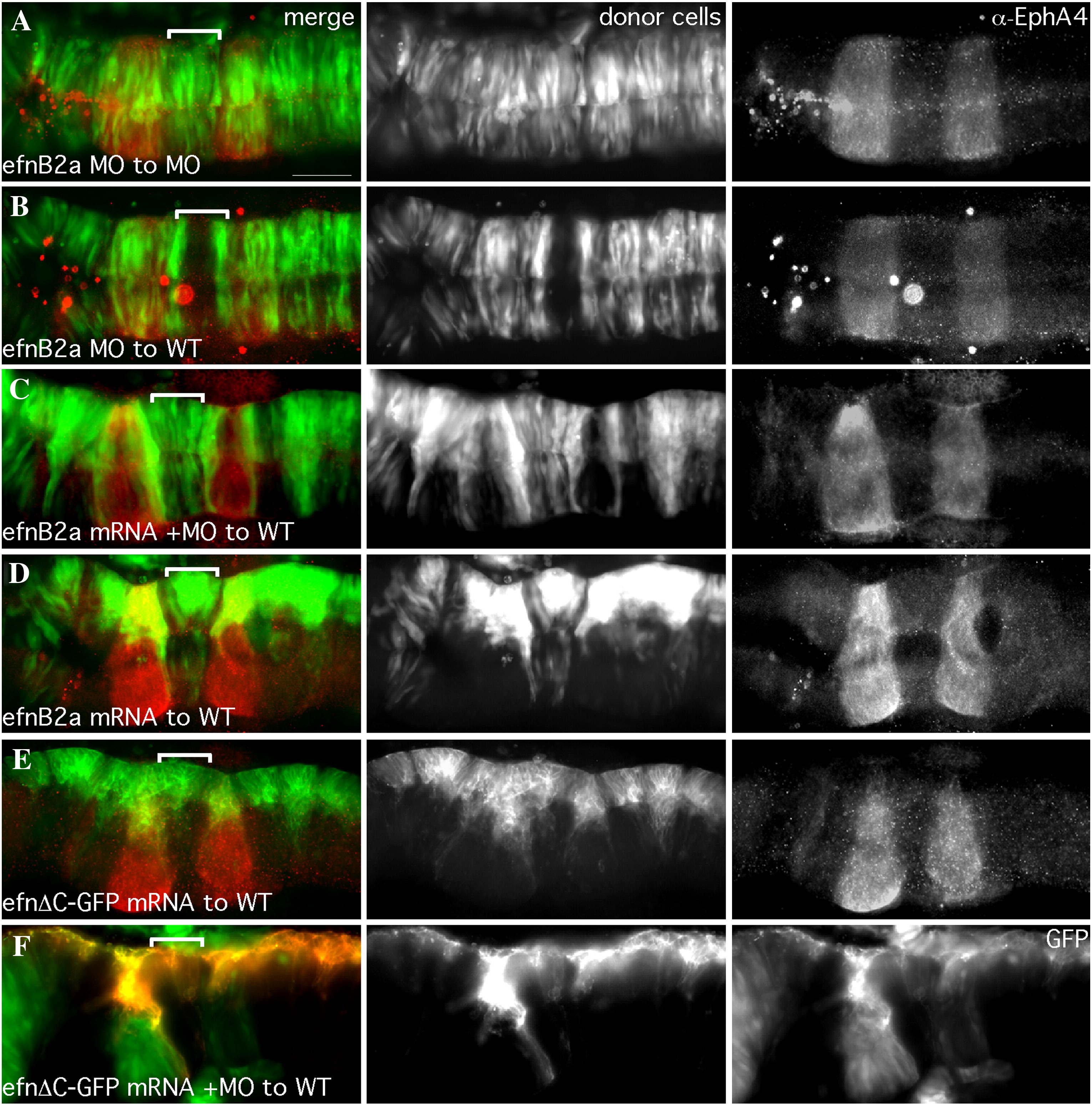
Fig. S1 efnB2a mRNA expression rescues cell?cell adhesion in EfnB2a MO r4, while over-expression induces adhesion-based clustering. Dorsal views of 18 hpf embryos with anterior to the left. r4 is indicated by a white bracket. Left hand panels are merged images of individual channels. Middle panel: Donor cells, pseudocolored in green (A?E) or red (F) in the merge, express GFP (A?E) or memb-RFP (F) from injected mRNA. r3 and r5 are visualized by α-EphA4 staining (red), or by the expression of GFP in r3 and r5 of a transgenic pGFP5.3 host. (A) Cells depleted of Efnb2a by MO injection contribute normally to an EfnB2a MO host embryo. (B) Cells depleted for EfnB2a by MO are excluded from r4 of a WT host. (C) EfnB2a MO donor cells coinjected with EfnB2a mRNA contribute to r4 of WT hosts. Ectopically expressing EfnB2a+ donor cells are simultaneously excluded from rhombomeres that do not normally express EfnB2a and instead express Eph receptors (r2,3,5, and r6). (D) WT donor cells injected with a ?rescuing? amount of EfnB2a mRNA form unilateral cell clusters in r4 of a WT host. (E) WT donor cells expressing the EfnB2aΔC-GFP allele, in which the C-terminus is replaced with GFP, fail to cross the midline in r4 of a WT host. Note that this GFP fusion protein is localized to cell membranes, as expected. (F) Efnb2a MO donor cells expressing the EfnB2aΔC-GFP allele also fail to cross the midline in r4 of a WT host. Note that donor cells are visible in the GFP channel, confirming expression of the truncated allele. Scale bar: 50 μm.
Reprinted from Developmental Biology, 327(2), Kemp, H.A., Cooke, J.E., and Moens, C.B., EphA4 and EfnB2a maintain rhombomere coherence by independently regulating intercalation of progenitor cells in the zebrafish neural keel, 313-326, Copyright (2009) with permission from Elsevier. Full text @ Dev. Biol.

