
|
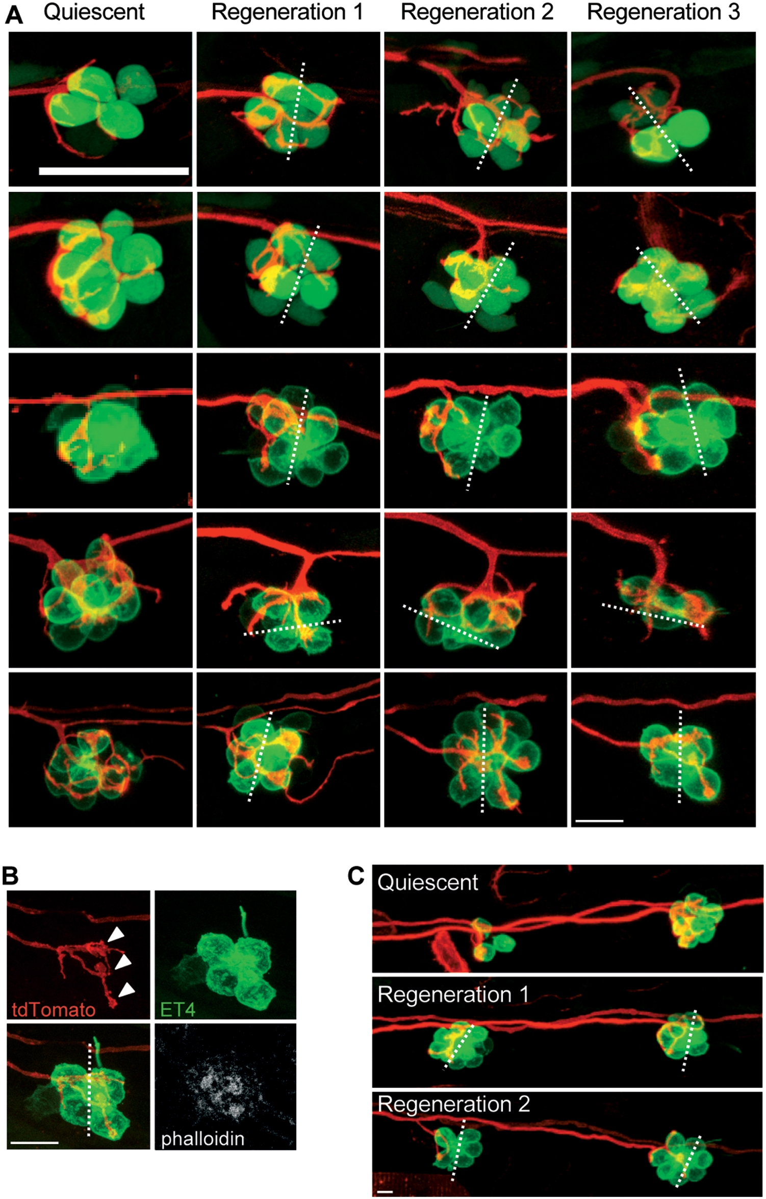
Fig. 6 Afferent neurons reinnervate the hair cells of the same polarity after several cycles of ablation and regeneration.
(A) Maximal projections of SqET4 (two first lines) or brn3c:GFP fish neuromasts innervated by a single neuron expressing mem-TdTomato. Confocal z-stacks of the same neuromast have been acquired at 3 dpf (quiescent state, first row) and 20 hours after each of three rounds of neomycin treatments (Regeneration 1, 2 and 3). All rows picture neuromasts with parallel cellular polarity (hair bundles aligned with the anteroposterior axis of the fish body) except the penultimate, which shows a neuromast with perpendicular cellular polarity. (B) Phalloidin staining performed on the last neuromast depicted on A after the third regeneration process. (C) Maximal projections of two brn3c:GFP adjacent neuromasts innervated by a single neuron expressing mem-TdTomato. Confocal z-stacks of the same neuromasts have been acquired at 3 dpf (quiescent) and 20 hours after each of two rounds of neomycin treatments (Regeneration 1 and 2). Dotted lines represent the axis of polarity of the neuromast. White arrowheads in panel B indicate the bulged neurites establishing contacts with the two hair cells of same polarity. Scale bar: 10 μm.