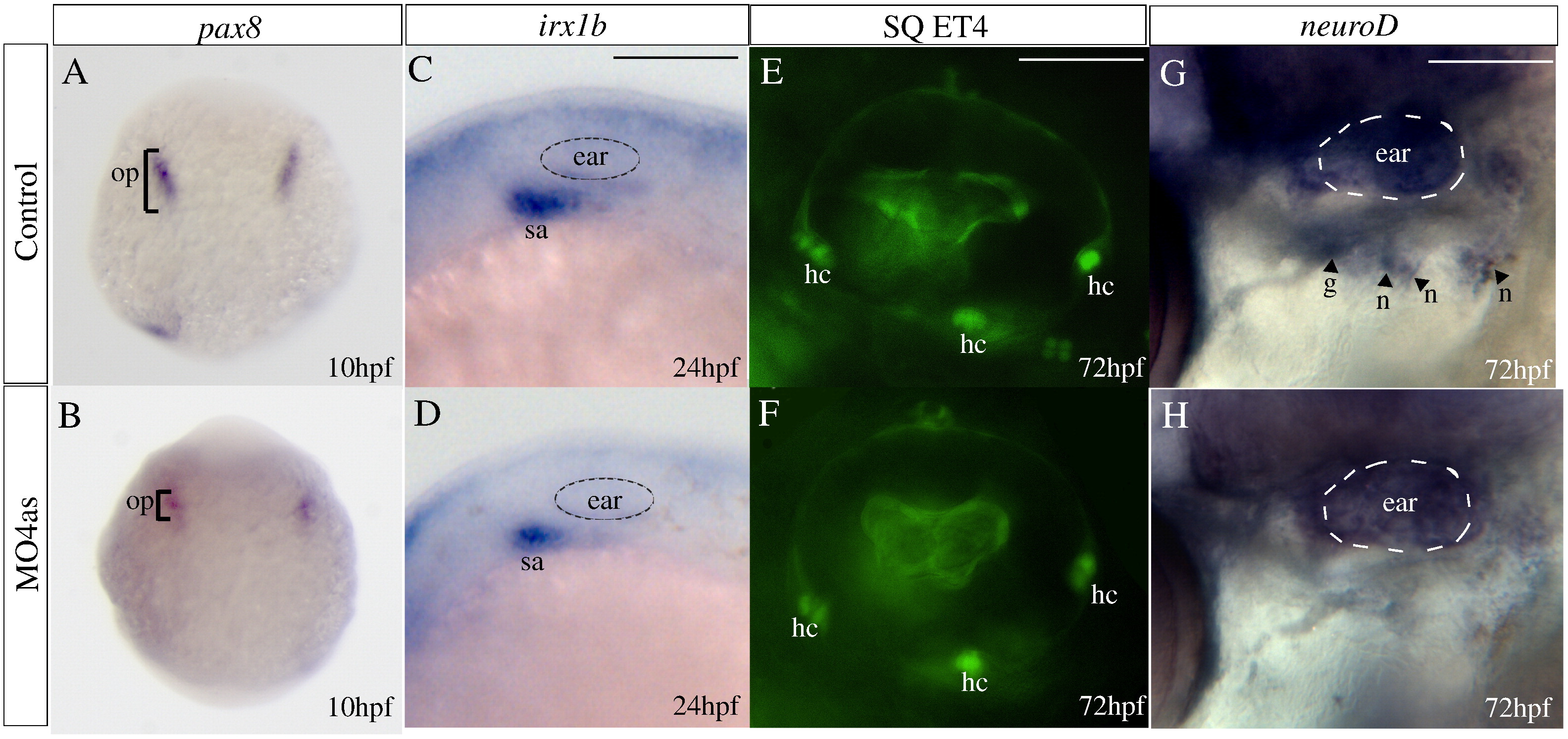
Fig. 5 Requirement of irx4a for epibranchial ganglia but not ear development. In situ hybridization results (A?D, G?H) and images of GFP expression in transgenic larvae (E?F) for control and irx4a morphant fish. (A, B) Expression of the otic placode marker pax8 is slightly decreased in irx4a morphant embryos at 10 hpf. (C, D) Expression of the statoacoustic ganglion marker irx1b is similarly affected at 24 hpf. (E, F) Examination of hair cells (hc) in control and morphant SqET4 transgenic larvae shows no effect of the morpholino injection at 72 hpf. In this transgenic line, the three patches of hair cells in the ear can be observed in live fish. (G?H) In situ hybridization showing neuroD expression in the epibranchial ganglia (petrosal, p, and nodose, n) at 72 hpf. Note the absence of expression of this marker in morphant (H) compared to controls (G). hc, clusters of hair cells; n, nodose ganglia; sa, statoacoustic ganglion; o, otic placode; p, petrosal ganglion. A, B dorsal views of whole embryos. C?H lateral views centered on the otic vesicle. Scale bar in C: 15 μm for C, D; scale bar in E: 5 μm for E, F; scale bar in I: 10 μm for G, H.
Reprinted from Molecular and cellular neurosciences, 40(3), Feijoo, C.G., Saldias, M.P., De la Paz, J.F., Gómez-Skarmeta, J.L., and Allende, M.L., Formation of posterior cranial placode derivatives requires the Iroquois transcription factor irx4a, 328-337, Copyright (2009) with permission from Elsevier. Full text @ Mol. Cell Neurosci.

