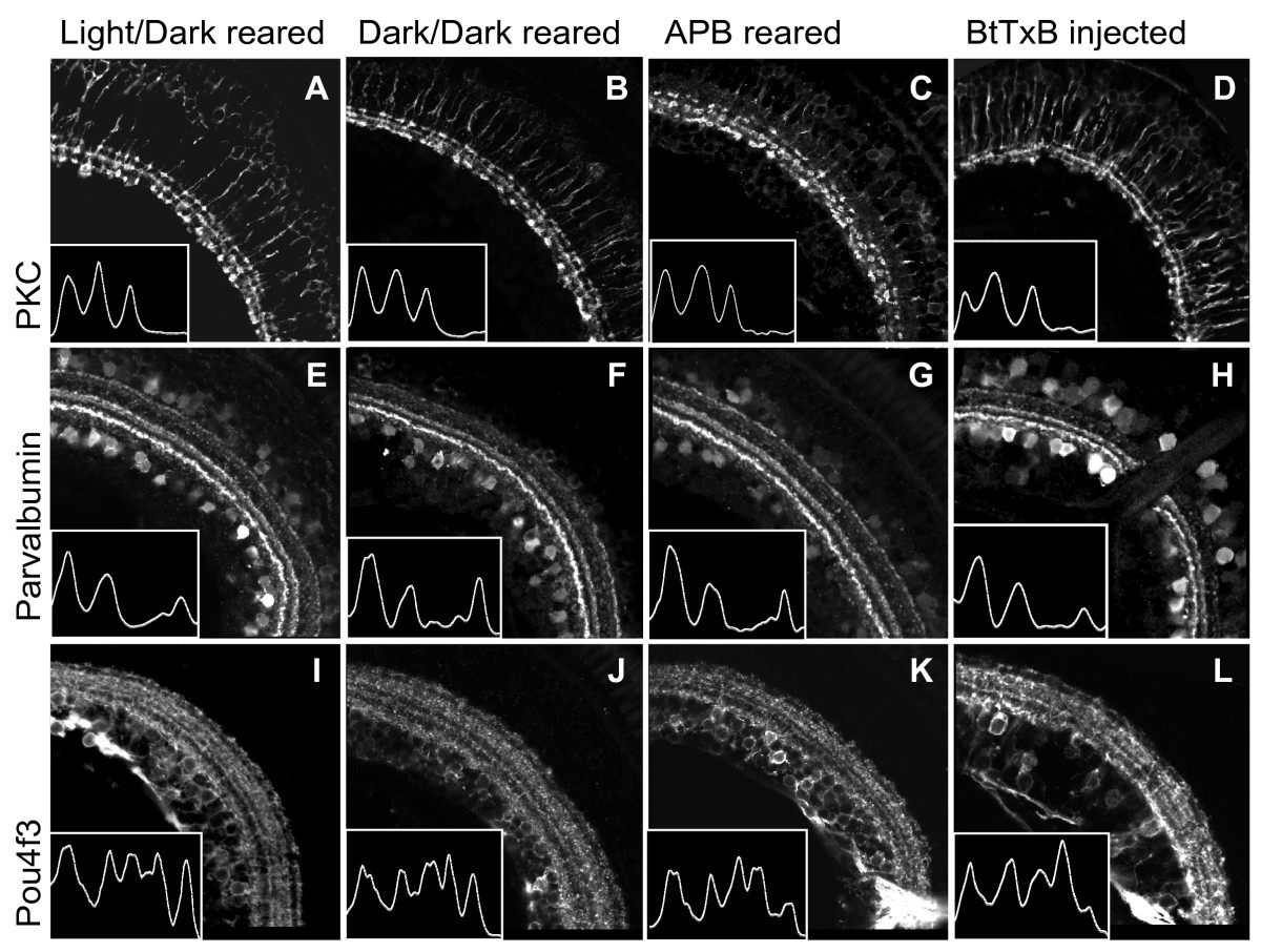
Fig. 3 Dark-reared, APB-treated, and BtTxB-injected larvae show proper IPL sublamination.(A-L) Sections showing the IPL of 5 dpf larvae raised in a normal light:dark cycle (A, E, I), constant darkness (B, F, J), in the presence of 1 mM APB (C, G, K), and treated with BtTxB (D, H, L). The images in D, H, L are from the larva recorded in Figure 4D. Insets: traces of the fluorescent signal intensity across the width of the IPL (region shown). Peaks correspond to bands in the IPL. (A-D) PKC+ BC axon terminals are confined to three inner sublaminae in all larvae. (E-H) Parv+ neurites are in three bands in all larvae. The interruption of the IPL in H is the optic nerve. (I-L) Pou4f3:mGFP+ dendrites stratify in five bands in all larvae. Scale bar 50 μm.
Image
Figure Caption
Acknowledgments
This image is the copyrighted work of the attributed author or publisher, and
ZFIN has permission only to display this image to its users.
Additional permissions should be obtained from the applicable author or publisher of the image.
Full text @ Neural Dev.

