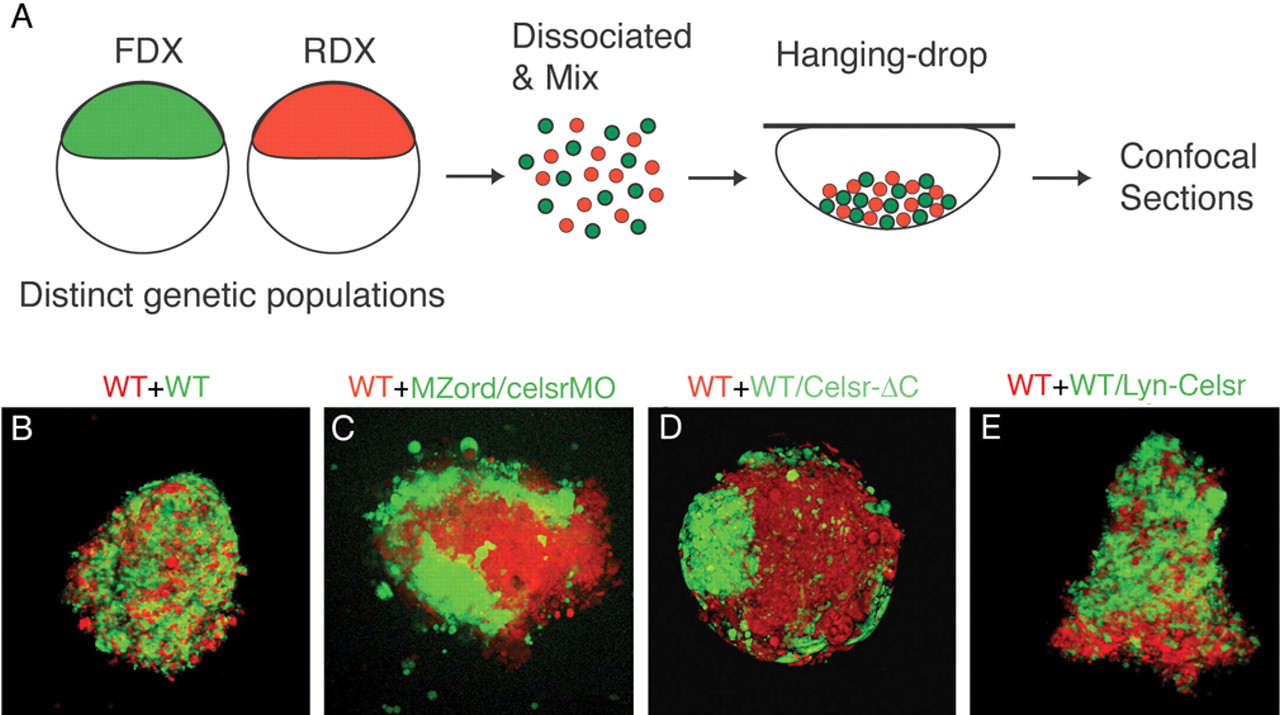
Fig. 7 Differential cell cohesive properties of wild-type cells compared with cells from celsr mutant/morphants and cells from embryos expressing Celsr-ΔC. Wild-type and MZord embryos were injected with RNA/morpholinos together with fluorescein-dextran (FDX, green) or rhodamine-dextran (RDX, red), as indicated. (A) 30-40% epiboly embryos were dissociated and dissociated cells from different populations were mixed in a minimal volume of a hanging drop and kept overnight to analyse aggregates by confocal microscopy. (B) Cells from wild-type embryos (green) and wild-type embryos (red). (C) Cells from MZord embryos injected with 0.4 pmoles each of celsr1a and celsr1b morpholinos (green) and wild-type embryos (red). (D) Cells from wild-type embryos injected with 300 pg Celsr-ΔC-HA RNA (green) and wild-type embryos (red). (E) Cells from wild-type embryos injected with 100 pg Lyn-Celsr RNA (green) and wild-type embryos (red). Celsr mutant/morphant cells (C) or Celsr-ΔC-expressing cells (D) are strongly segregated from wild-type cells, whereas Lyn-Celsr-expressing cells are only weakly segregated from wild-type cells (E).
Image
Figure Caption
Figure Data
Acknowledgments
This image is the copyrighted work of the attributed author or publisher, and
ZFIN has permission only to display this image to its users.
Additional permissions should be obtained from the applicable author or publisher of the image.
Full text @ Development

