
Image
|
Figure Caption
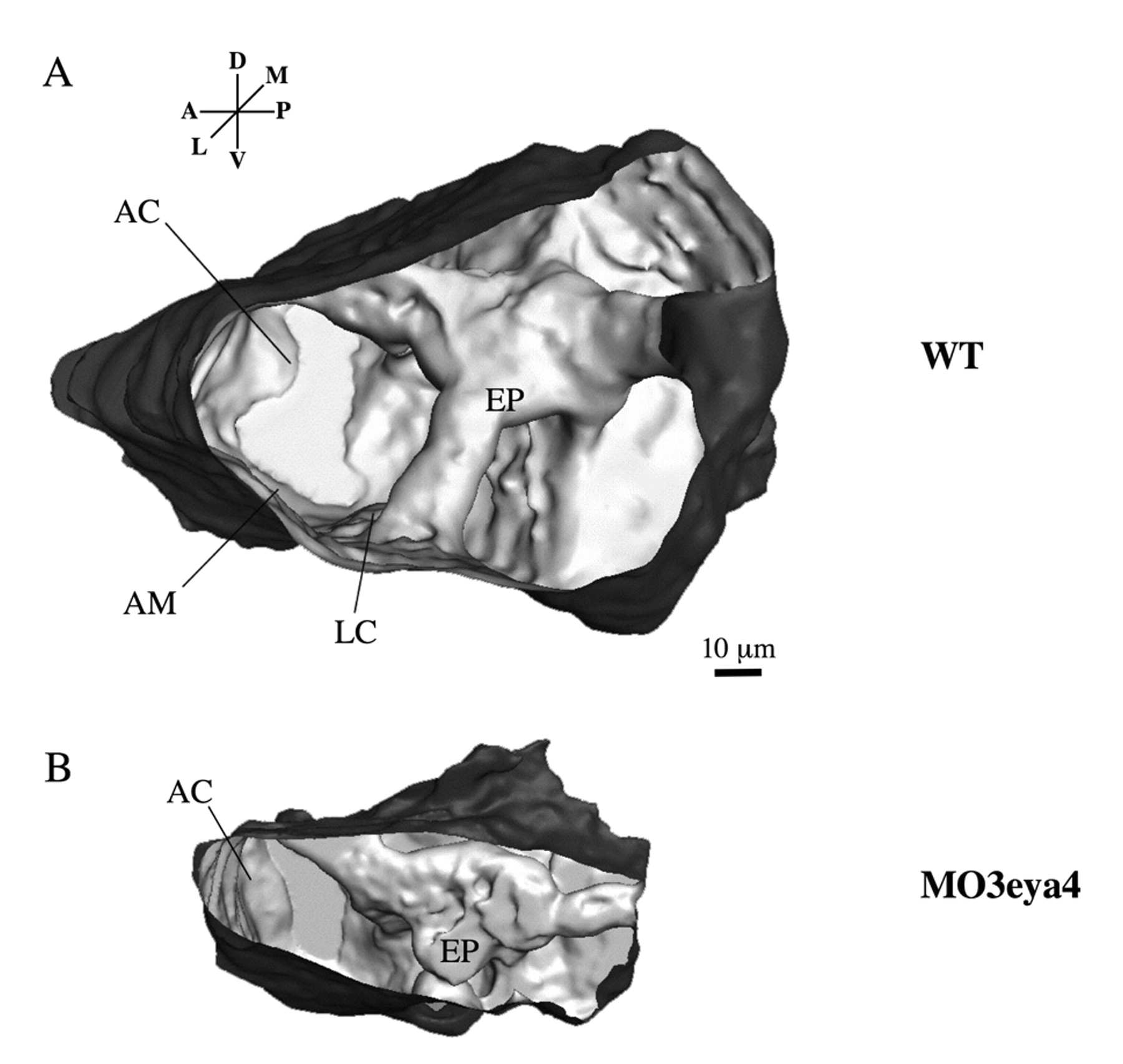
Fig. 2 Three-dimensional reconstruction of the zebrafish otic vesicle (72 hpf) generated from confocal images. (A) The otic vesicle of the wild-type zebrafish has well-formed epithelial pillars (EP) that shape the semicircular canals. The anterior cristae (AC), lateral cristae (LC) and anterior maculae (AM) are indicated. (B) The otic vesicle of an eya4 morphant fish is smaller and misshapened, with fused epithelial pillars and malformed semicircular canals.
Figure Data
Acknowledgments
This image is the copyrighted work of the attributed author or publisher, and
ZFIN has permission only to display this image to its users.
Additional permissions should be obtained from the applicable author or publisher of the image.
Full text @ Development