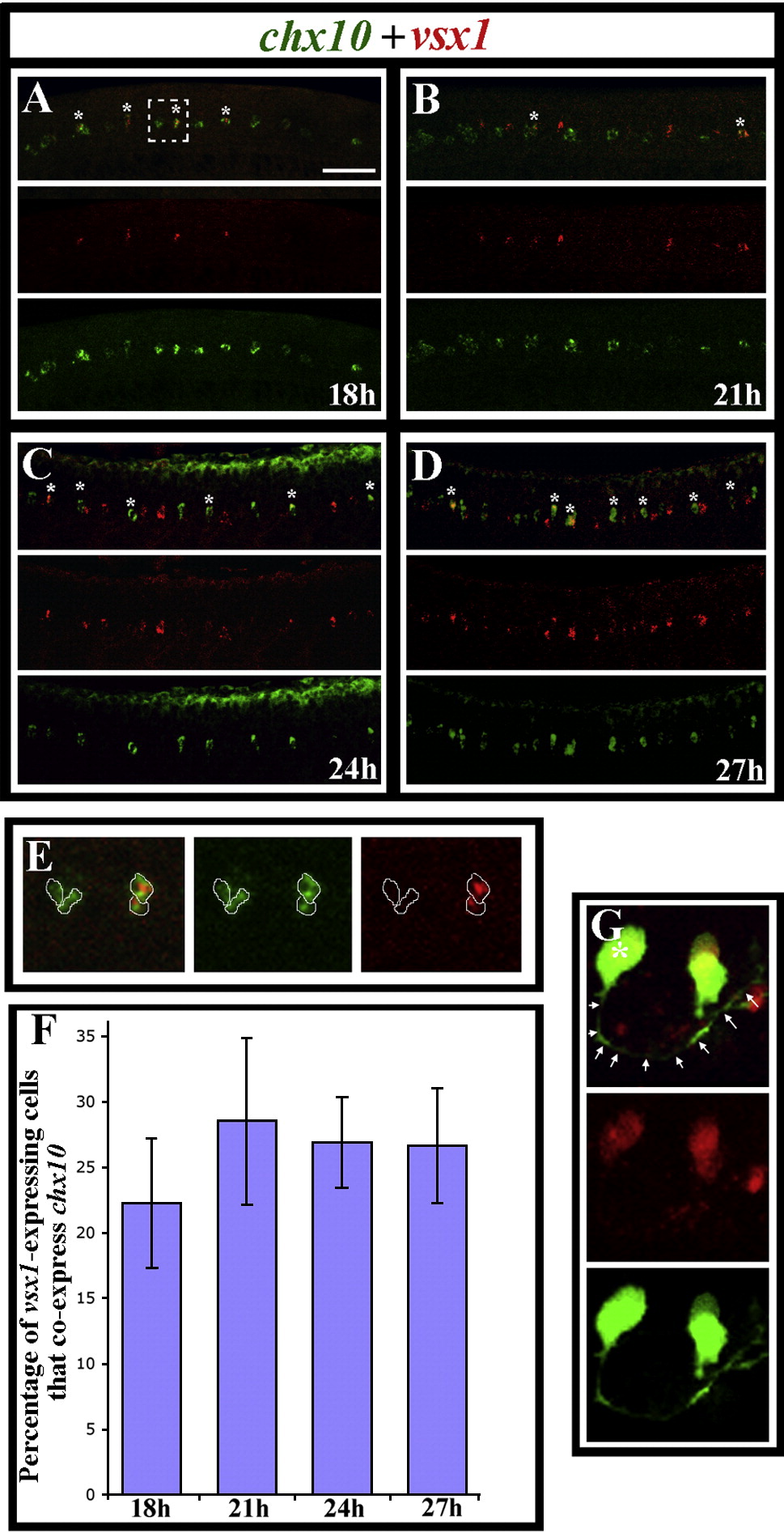
Fig. 4 vsx1 and chx10 are co-expressed by subsets of vsx1- and chx10-expressing cells. Lateral views of the spinal cord in wild-type trunks at (A) 18 h (18 somites), (B) 21 h, (C) 24 h and (D) 27 h. Double in situ hybridisations showing chx10 expression in green and vsx1 expression in red. Single channel views and merged images are shown. Anterior is left, dorsal is up. Scale bar = 50 μm (A?D). Double-labelled cells are indicated with stars. Panel E shows single focal planes of a higher magnification view of the white dashed square in panel A. Merged image is on the left, followed by the single green channel and then the single red channel. Cell outlines are traced in white. The 2 cells on the left only express chx10; the 2 cells on the right express both chx10 and vsx1. Panel F shows the percentage of vsx1-expressing cells that co-express chx10 at different stages. Cell counts are from a 5 somite length of spinal cord adjacent to somites 6?10. Values shown are the mean from 5 different embryos. Error bars indicate standard deviations. Panel G shows a vsx1-expressing cell with a clear CiD morphology in a 24 h Tg(chx10:Kaede) embryo. Single channel views and a merged image are shown. vsx1-expression is red. Kaede-positive cells are green. Star indicates cell body and arrows indicate the associated axon.
Reprinted from Developmental Biology, 322(2), Batista, M.F., Jacobstein, J., and Lewis, K.E., Zebrafish V2 cells develop into excitatory CiD and Notch signalling dependent inhibitory VeLD interneurons, 263-275, Copyright (2008) with permission from Elsevier. Full text @ Dev. Biol.

