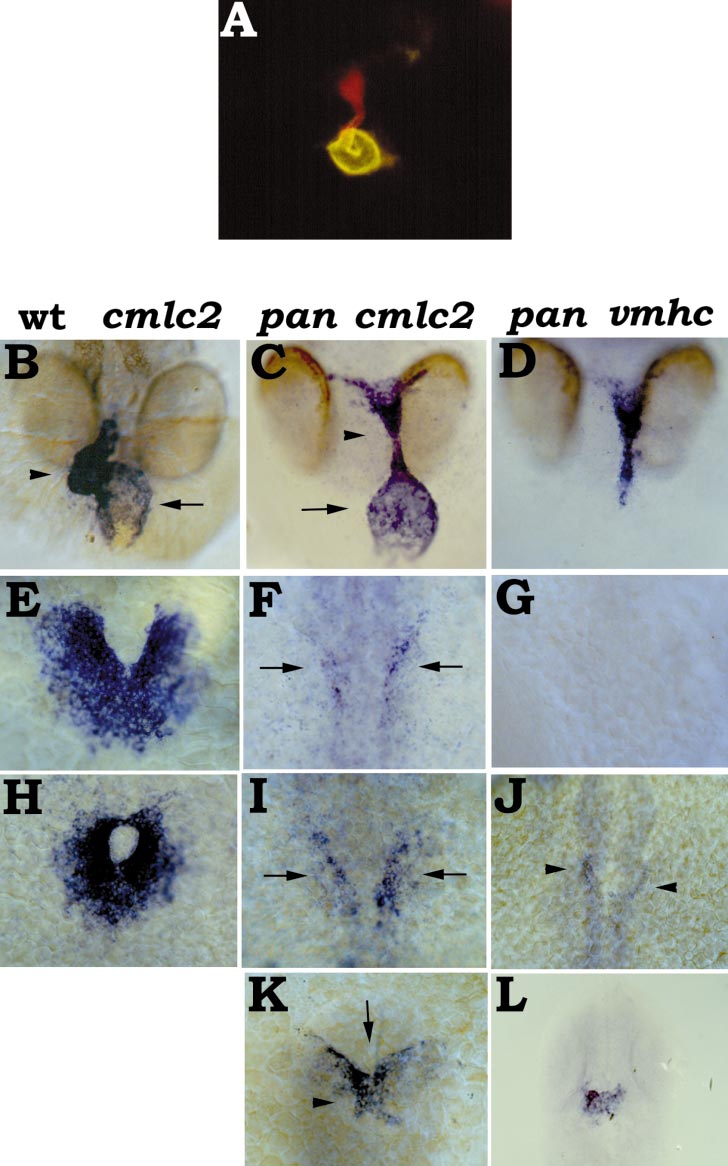
Fig. 7 The Pandora (pan) mutation affects ventricle formation as well as early expression of cmlc2 and vmhc. (A) Head-on view of a 48-hpf pan mutant embryo stained with MF20 (TRITC) and S46 (FITC), dorsal at the top. A thin stalk of ventricular tissue (red) is attached to the rostral end of a bulbous atrium (yellow). (B, E, H) Expression of cmlc2 in wild-type embryos. (C, F, I, K) Expression of cmlc2 in pan mutant embryos. (D, G, J, L) Expression of vmhc in pan mutant embryos. (B–D) Head-on views, dorsal at the top, of 48-hpf embryos. In pan mutants, the heart is composed of a bulbous atrium (C, arrow) and a thin stalk of ventricular tissue (C, arrowhead). (E–L) Dorsal views, anterior at the top. (E, F, G) 18-somite stage; pan embryos exhibit relatively faint and thin bilateral stripes of cmlc2-expressing cells (F, arrows), in contrast to the robust cmlc2-expressing population of fusing myocardial precursors in wild-type siblings (E). pan embryos do not express vmhc at this stage (G). (H, I, J) 21-somite stage; pan embryos exhibit slightly stronger cmlc2 expression (I, arrows), but still far less than in wild-type embryos (H). Some pan embryos have faint bilateral patches of vmhc expression (J, arrowheads). (K, L) 36 hpf; cardiac cone formation is delayed and abnormal in pan mutants (K). Often, pan mutant cones have a split base (K, arrow). The apex of the pan cone (K, arrowhead) expresses a significant amount of vmhc (L). It is important to note that pan embryos lag behind their wild-type siblings during somitogenesis. For example, pan embryos usually have only 16 somites when their wild-type siblings have 20 somites. In (E–G) and (H–J), we are comparing pan embryos with wild-type embryos with the same number of somites; i.e., the wild-type siblings are fixed a few hours before the pan mutants. Also note that the precise morphology of the developing myocardium in pan mutants can vary; these examples represent typical expression patterns observed in a majority of mutants.
Reprinted from Developmental Biology, 214(1), Yelon, D., Horne, S.A., and Stainier, D.Y.R., Restricted expression of cardiac myosin genes reveals regulated aspects of heart tube assembly in zebrafish, 23-37, Copyright (1999) with permission from Elsevier. Full text @ Dev. Biol.

