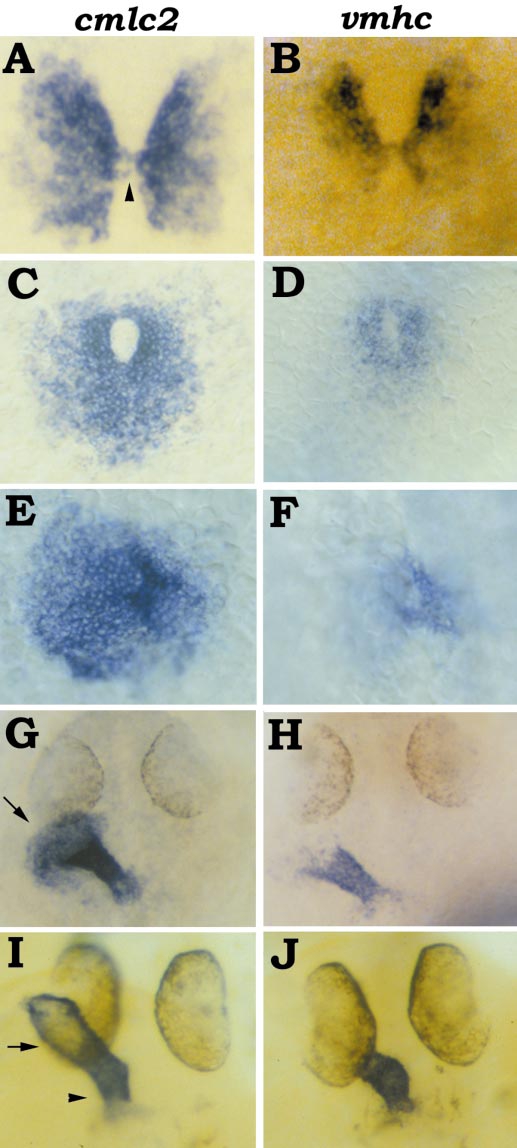
Fig. 4
Following cmlc2 and vmhc expression during heart tube assembly. (A, C, E, G, I) Expression of cmlc2. (B, D, F, H, J) Expression of vmhc. All panels show dorsal views, anterior at the top. (A, B) 18-somite stage; the myocardial precursors (A) make contact via a bridge (arrowhead) of vmhc-expressing cells (B). (C, D) 21-somite stage; the cardiac cone (C) forms with vmhc-expressing cells (D) at its center and apex. (E, F) 23-somite stage; the cardiac cone (E) begins its transformation into a tube with the extension and tilting of the
Reprinted from Developmental Biology, 214(1), Yelon, D., Horne, S.A., and Stainier, D.Y.R., Restricted expression of cardiac myosin genes reveals regulated aspects of heart tube assembly in zebrafish, 23-37, Copyright (1999) with permission from Elsevier. Full text @ Dev. Biol.

