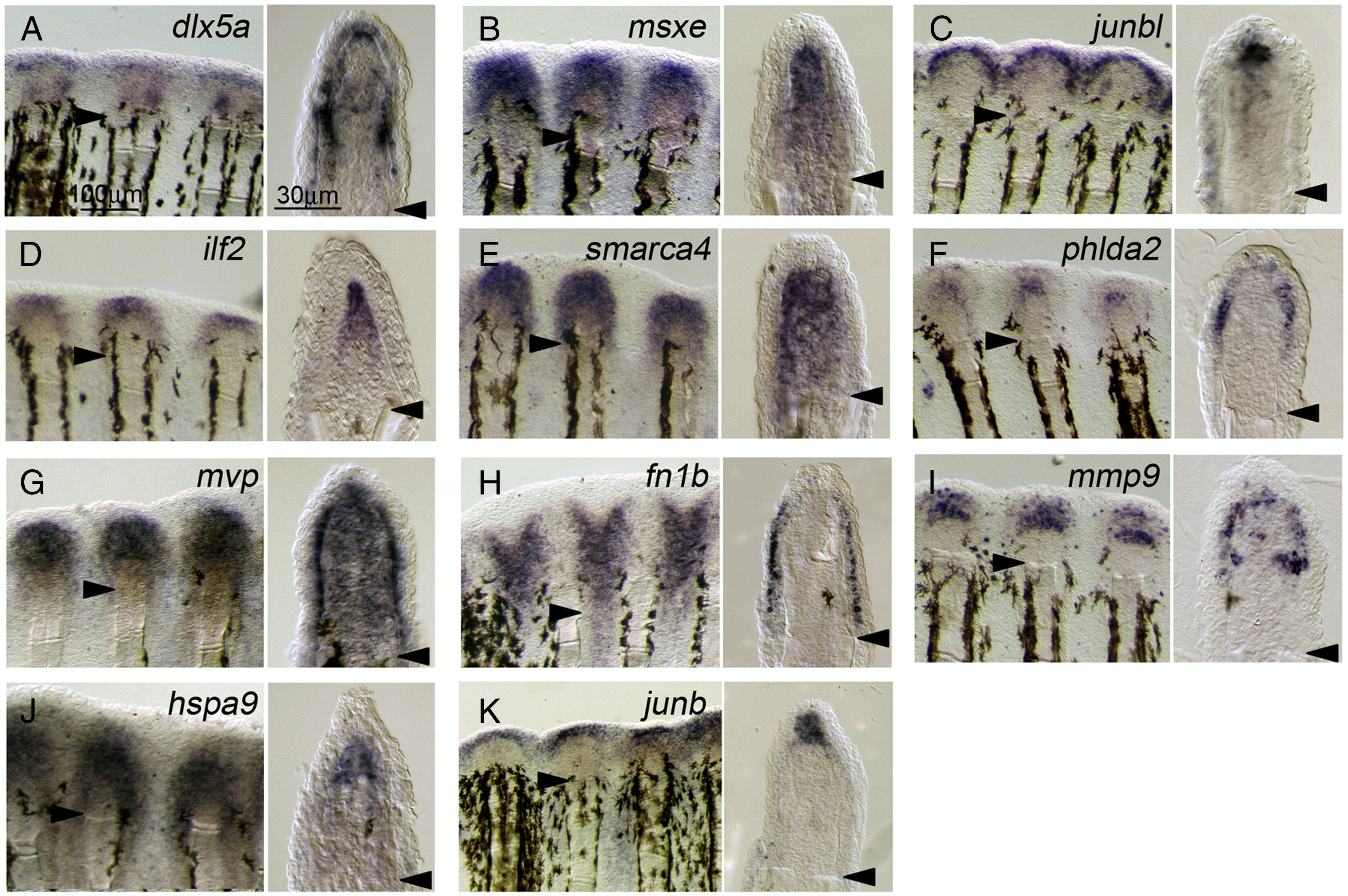
Fig. 3
Fig. 3 Localized expression of regeneration-related transcripts during adult regeneration. (A?K) ISH analysis showing the respective gene expression at 2 dpa of adult regeneration. Respective gene expressions were detected in whole-mount preparations (left panels), and their cellular localizations were assessed in sections (right panels). The expression of dlx5a (A) had 3 domains: the basal wound epidermis, distal blastema, and cells at proximal and lateral regions of the blastema. The expressions of msxe (B), ilf2 (D), smarca4 (E), and hspa9 (J) were seen exclusively in the blastema, whereas junbl was only in the distal part of blastema (C). Similarly, the wound epidermis can be subdivided into several distinct regions: the lateral part of basal wound epidermis expressing phlda2 (F), the basal wound epidermis expressing mvp (G), a layer of regenerating epidermis expressing fn1b (H), and the apical epidermis expressing junb (K). Furthermore, mmp9 expression was seen in cells irregularly distributed within the basal wound epidermis and in cells at the proximal and lateral region of the blastema, which appear to overlap with those expressing dlx5a. As in the larval fin fold, the expression of all genes was not detected in uncut fins. Arrowheads mark the sites of amputation. The same magnifications for respective right and left columns of panels A?K (scale bars in panel A).
Reprinted from Developmental Biology, 325(1), Yoshinari, N., Ishida, T., Kudo, A., and Kawakami, A., Gene expression and functional analysis of zebrafish larval fin fold regeneration, 71-81, Copyright (2009) with permission from Elsevier. Full text @ Dev. Biol.

