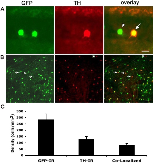
Fig. 3 Double immunostaining using anti-GFP and anti-TH antibodies. Immunostaining experiments were performed in vertical retinal sections (A) and the whole-mount retina (B). GFP-IR, TH-IR, and GFP-IR/TH-IR are shown in green, red and yellow, respectively. Quantification of double-staining in the whole-mount retina is shown in C. Values represent the mean±SD from 4 individual retinas. The cell density was calculated by dividing the number of cells by the image area calculated by MetaMorph. Overall, 29±2% of GFP-labeled cells coexpressed TH. Scale bar equals 10 μm for A and 20 μm for B. Arrowhead in A points to a GFP-IR/non-TH-IR cell. Arrowhead in B points to a TH-IR/non-GFP-IR cell.
Image
Figure Caption
Figure Data
Acknowledgments
This image is the copyrighted work of the attributed author or publisher, and
ZFIN has permission only to display this image to its users.
Additional permissions should be obtained from the applicable author or publisher of the image.

