
|
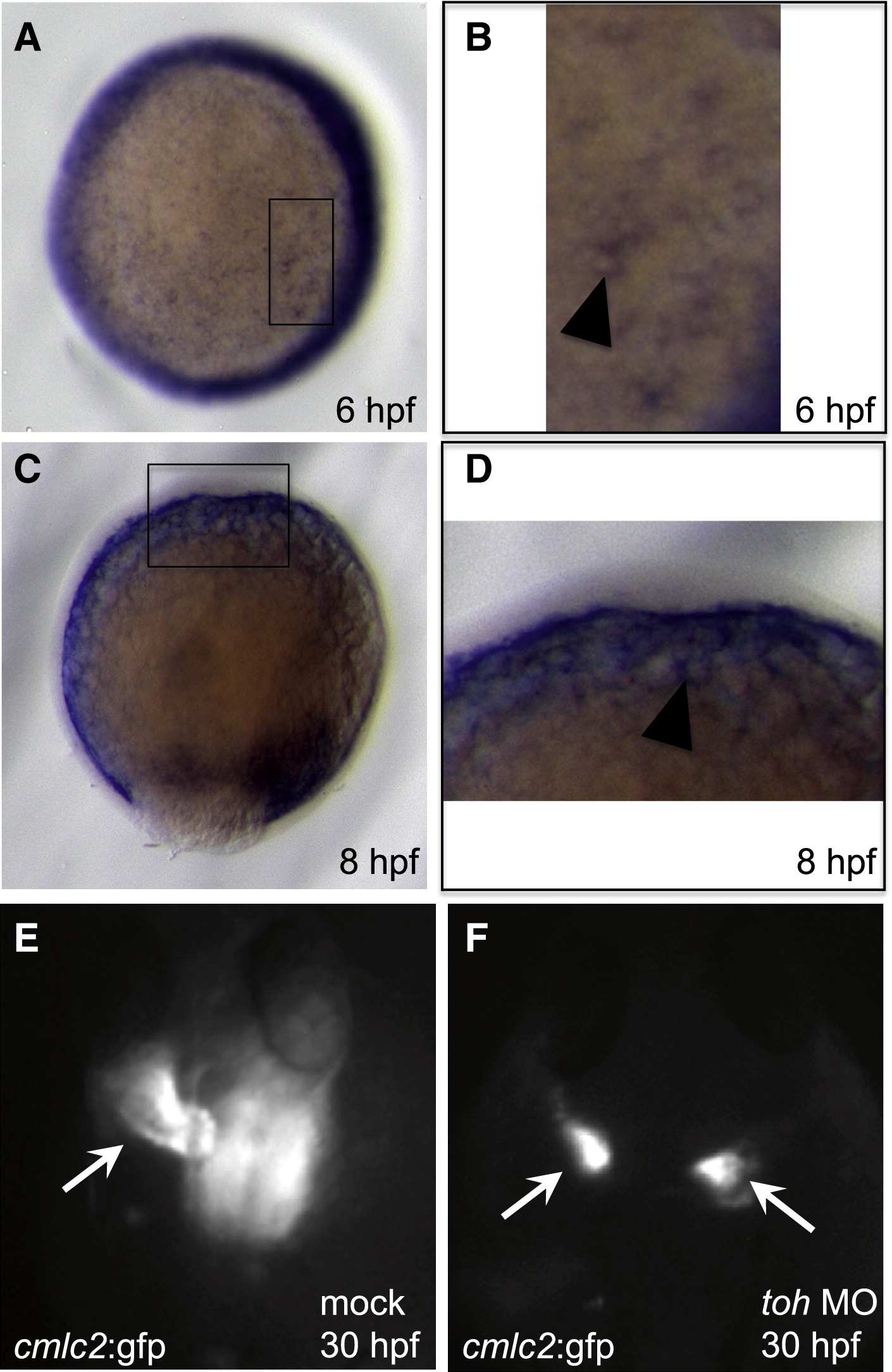
Fig. 3 Toh Function in the YSL Is Required for Precardiac-Mesoderm Migration
(A?D) toh expression at 6 (A and B) and 8 (C and D) hpf. (A) Animal-pole view with dorsal to the right, 6 hpf, showing toh expression around the margin and diffusely throughout the YSL. (B) Magnified view of the box in (A) showing toh expression around the YSL nuclei (arrowhead). (C) Lateral view with dorsal to the right, 8 hpf, showing continued toh expression in cells that have involuted, as well as in the YSL. (D) Magnified view of the box in (C) showing pronounced toh expression in the YSL (arrowhead).
(E and F) Dorsal images of Tg(cmlc2:GFP)f1 embryos injected into the YSL with mock solution (E) or with 4 ng toh MO (F) and visualized at 30 hpf. Mock-injected embryos have a single heart tube (E, arrow), whereas embryos with loss of Toh function in the YSL very frequently (84%, n = 56) display cardia bifida (F; arrows).