Fig. 3
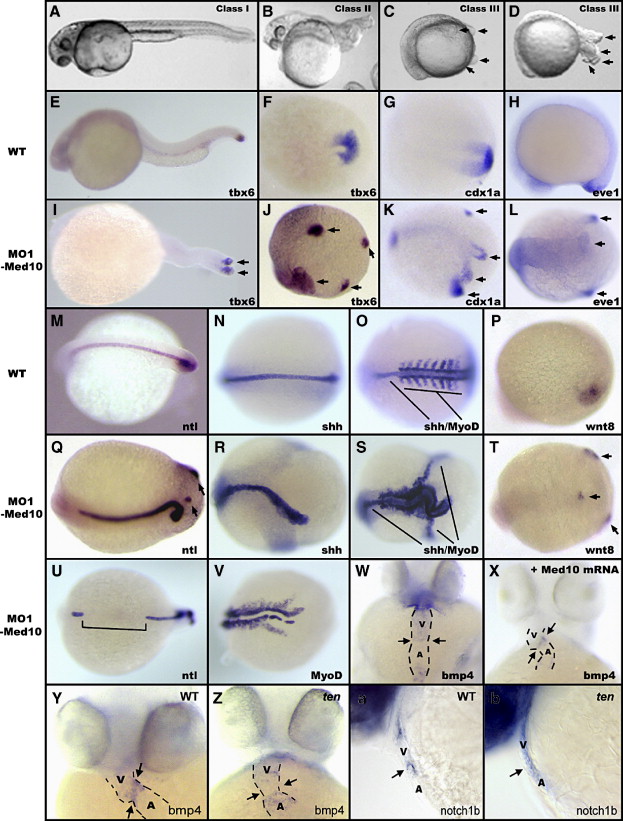
Fig. 3 Reduction of Med10 results in specific developmental defects in a dose-dependent manner. (A–D) Reduction of Med10 leads to three classes of defects: (A) Class I embryos, (B) Class II embryos, (C) Class III embryos. The tails in approximately 14% (12/88) of Class III embryos later extend out from the body and clearly exhibit multiple tail buds (D). (E–V) Injection of high dosages of MO1-Med10 (15 ng) leads to altered notochord extension and tail bud formation. In situ hybridization was performed on 30 hpf injected embryos or the age-matched (by counting the somite number) control embryos. Two tail buds can be detected in Class II mutants, as shown by tbx6 staining (I). Approximately four tail buds formed in Class III embryos, as shown by tbx6 (J), cdx1a (K), eve1 (L), ntl (Q,U), and wnt8 (T) staining. The notochord in Class III embryos is indicated by ntl (Q,U) and shh staining (R,S). The somites in Class III embryos are indicated by MyoD staining (S,V). Arrows indicate positions of the multiple tail buds. Shown are dorsal views (F,G,J–O,Q–V) and lateral views (E,H–I,P) with anterior to the left. (W) Injection of low dosages of MO1-Med10 (5 ng) leads to whole-heart expression of bmp4, which can be rescued to the cardiac cushion-restricted expression pattern by co-injection with med10 mRNA (2 ng) (X). (Y-b) ten mutants exhibit cardiac cushion defects. Shown are bmp4 expression in wild-type (Y) and ten mutant (Z) embryos, and notch1b expression in wild-type (a) and ten mutant (b) embryos. Arrows indicate the atrial–ventricular cushion. Shown in (W–Z) are ventral views of 48 hpf embryos. Anterior is up. Shown in (a,b) are lateral views of 48 hpf embryos. Anterior is left and dorsal is up. V, Ventricle. A, Atrium.
Reprinted from Developmental Biology, 303(2), Lin, X., Rinaldo, L., Fazly, A.F., and Xu, X., Depletion of Med10 enhances Wnt and suppresses Nodal signaling during zebrafish embryogenesis, 536-548, Copyright (2007) with permission from Elsevier. Full text @ Dev. Biol.

