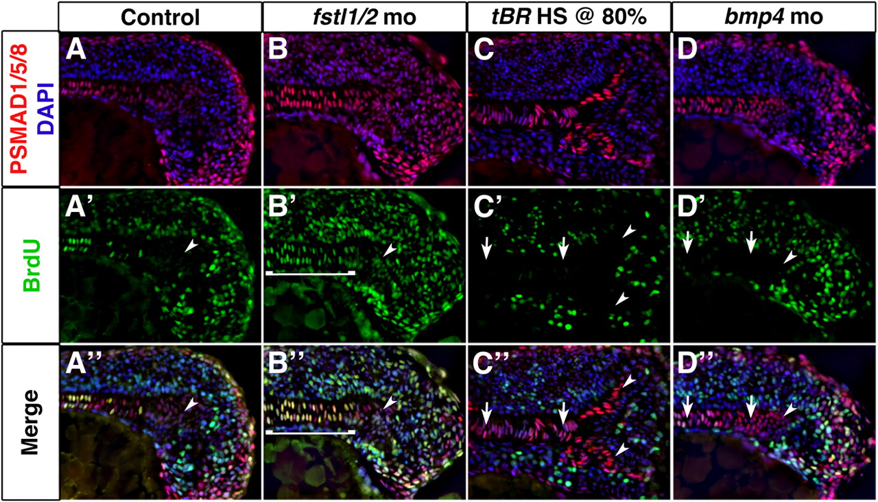
Fig. 4 BMP activity establishes proliferation in axial mesoderm. (A-D″) Longitudinal sections through the tailbud of 14-somite embryos exposed to PSMAD1/5/8 (red) and BrdU (green) antibodies reveal that axial cells undergoing proliferation are responding to BMP activity. Cell proliferation is increased in axial tissue of Fstl1/2 morphants (B-B″), and reduced in tBR embryos heat-shocked at 80% epiboly (C-C″) and Bmp4 morphants (D-D″). Arrowheads indicate the CNH. Anterior is towards the left in all views. Brackets and arrows indicate the expanse and absence of proliferation in chordamesoderm, respectively.
Image
Figure Caption
Figure Data
Acknowledgments
This image is the copyrighted work of the attributed author or publisher, and
ZFIN has permission only to display this image to its users.
Additional permissions should be obtained from the applicable author or publisher of the image.
Full text @ Development

