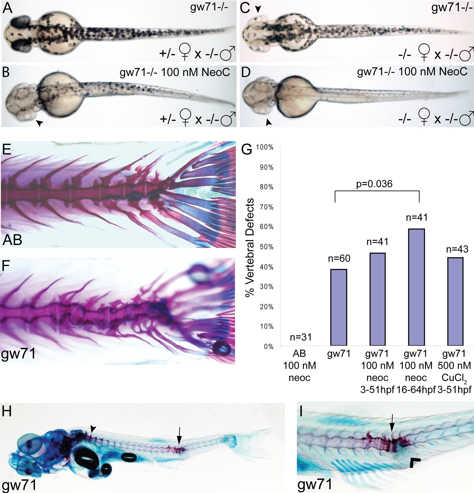
Fig. 3 calamitygw71 embryos display developmental defects that are sensitive to maternal and environmental copper availability.
(A?D) Maternal effect on pigmentation in untreated gw71 homozygous embryos. Mutant embryos derived from a heterozygous mother (A) display near normal pigmentation and have an incomplete loss of pigmentation in 100 nM neocuproine (B) most noticeable in the retina (arrowhead). Mutant embryos derived from homozygous mothers have mild hypopigmentation (C), particularly of the retina (arrowhead) and lose nearly all pigmentation in 100 nM neocuproine (D). (E?I) Partially penetrant juvenile skeletal deformities are present in gw71 mutant fish. Wild-type (E) and gw71 mutant (F) 21 dpf larvae were stained with alcian blue (cartilage) and alizarin red (bone) to reveal skeletal defects. In (G), embryos were untreated, treated with 100 nM neocuproine, or treated with 500 nM CuCl2 during the times indicated. The larvae at 21 dpf were scored according to absence or presence of a vertebral axis defect. A one-tailed Fisher exact probability test was used to calculate p-values. Only the indicated p-value was significant. (H) gw71 mutants at an earlier stage of bone ossification display hyperossification at the location of the vertebral defect (arrow). Normal ossification is detected by alizarin red staining and begins rostrally (arrowhead). (I) A higher magification of the defect in (H) showing the hyperossification (arrow) and an outpouching of connective tissue which stains with alcian blue (arrowhead).

