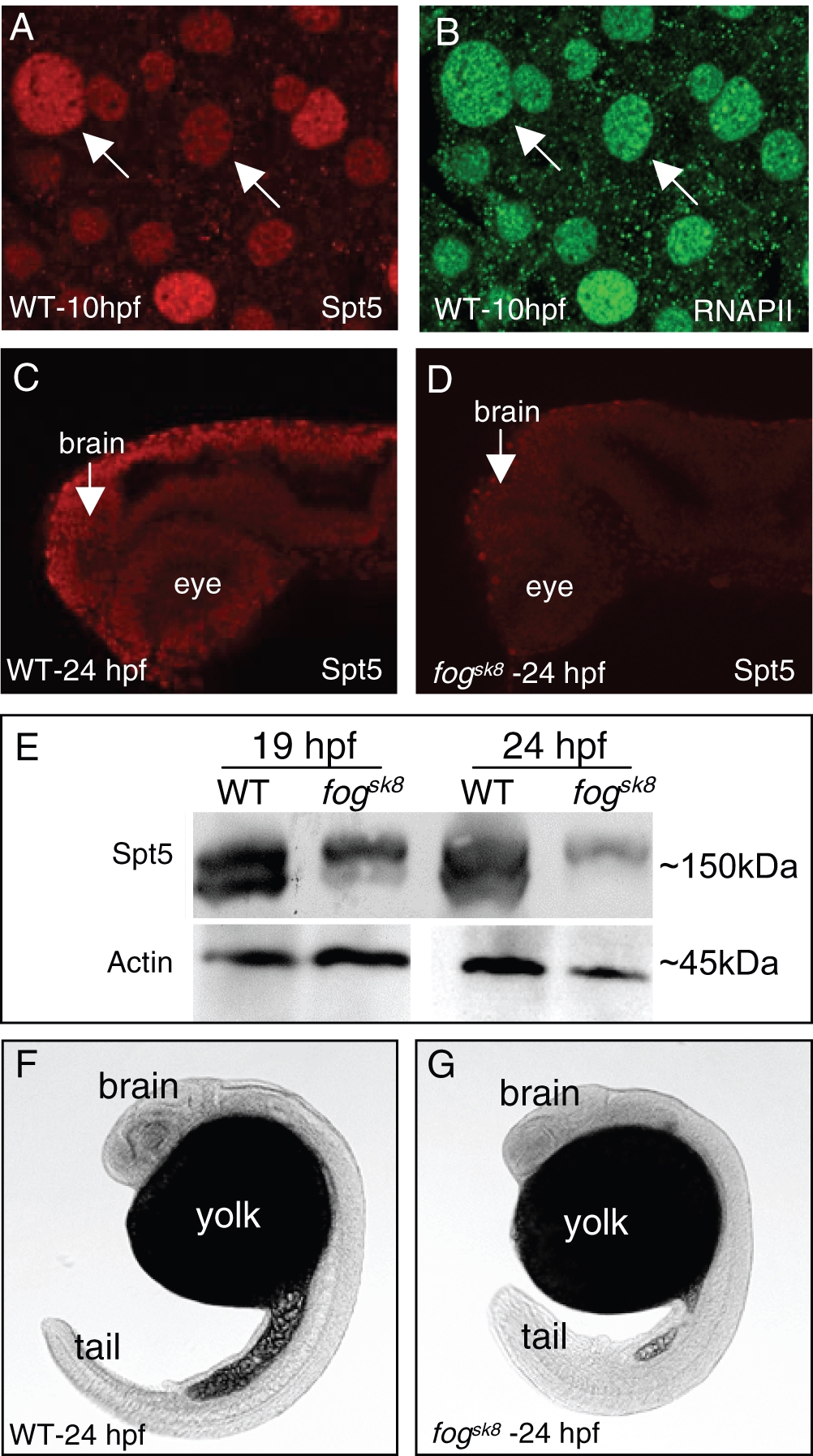
Fig. 1 Spt5 protein is significantly decreased in fogsk8 embryos.
(A?B) Whole mount immunofluorescence detection of Spt5 and RNAPII in zebrafish embryos, showing the nuclear localization pattern of Spt5 protein. (C?D) Whole mount immunofluorescence detection of Spt5 in WT and fogsk8 embryos at 24 hpf, showing overall decrease of Spt5 protein in fogsk8 embryos. Confocal images of lateral views are shown, with the anterior to the left and dorsal up. (E) Western blot of wild-type (WT) and fogsk8 embryonic extracts at 19 hpf and 24 hpf shows substantial decrease in maternal Spt5 protein in the mutant. (F?G) Light microscopic view of WT and sk8 embryos at 24 hpf, showing the gross morphological phenotype of the mutant embryos.

