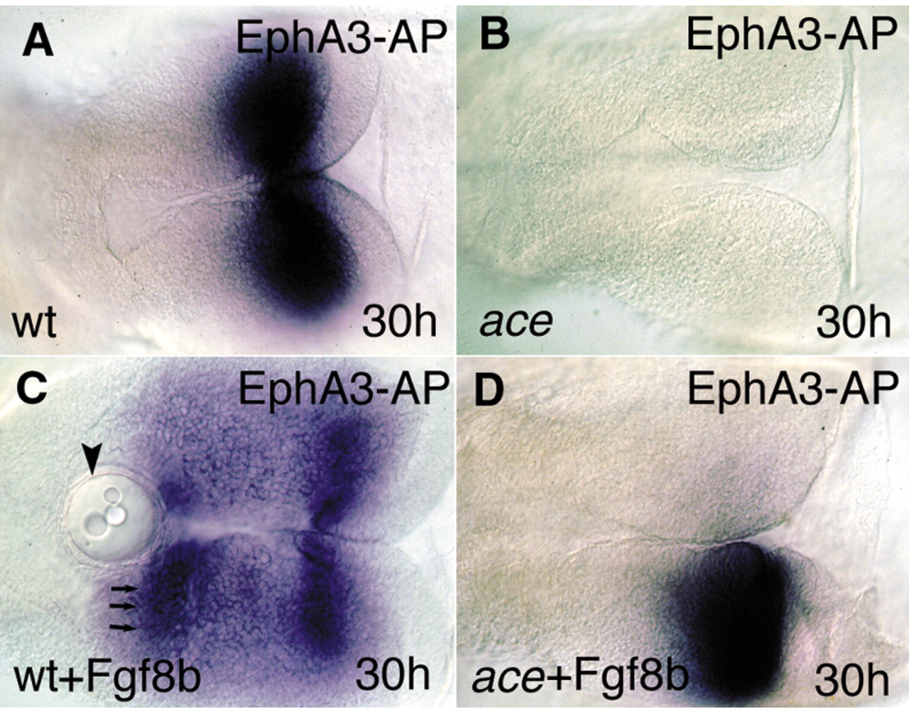
Fig. 7 Fgf8-bead implantation restores the molecular and anatomical identity of the MHB territory. All views are rostral to the left, and are dorsal aspects. (A) The Epha3-AP fusion protein reveals the distribution of ephrin ligands (blue) in the mesencephalic tectum. (B) The fusion protein fails to detect the typical ephrin gradient expression in the mutant tecta. (C) After implanting Fgf8-coated beads into the wild-type diencephalon, a second, mirror gradient of ephrin A proteins can be detected. Arrowhead indicates the implanted beads; arrows indicate the second, ectopic ephrin A domain. (D) Unilateral implantation of the Fgf8-coated beads into ace embryos is able to restore the graded ephrin A expression on the operated side.
Image
Figure Caption
Acknowledgments
This image is the copyrighted work of the attributed author or publisher, and
ZFIN has permission only to display this image to its users.
Additional permissions should be obtained from the applicable author or publisher of the image.
Full text @ Development

