Image
Figure Caption
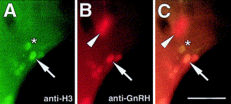
Fig. 5 H-GnRH cells undergo cell division after becoming GnRH-positive. (A–C) Ventral view of 56 h embryo processed for both anti-H3 and anti-GnRH antibodies. (A) H-GnRH cells are dividing (arrow) as are non-H-GnRH cells (asterisk). (B) H-GnRH cells (arrow) are positive for anti-GnRH antibody. TN-GnRH cells are evident out of the plane of focus (arrowhead). (C) Colabeling of the H-GnRH cells for the anti-H3 (arrow) and anti-GnRH antibodies. Scale bar: (A–C) 40 μm.
Acknowledgments
This image is the copyrighted work of the attributed author or publisher, and
ZFIN has permission only to display this image to its users.
Additional permissions should be obtained from the applicable author or publisher of the image.
Reprinted from Developmental Biology, 257(1), Whitlock, K.E., Wolf, C.D., and Boyce, M.L., Gonadotropin-releasing hormone (gnrh) cells arise from cranial neural crest and adenohypophyseal regions of the neural plate in the zebrafish, Danio rerio, 140-152, Copyright (2003) with permission from Elsevier. Full text @ Dev. Biol.

