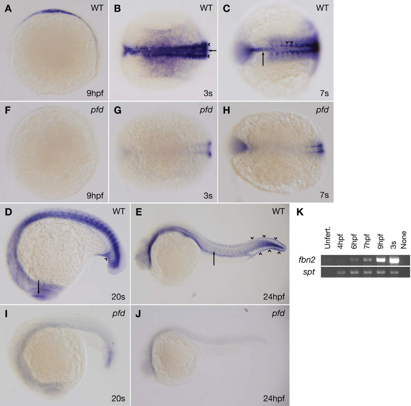
Fig. 7 fbn2 expression is consistent with the pfdgw1 phenotype and dramatically reduced in pfdgw1 mutants. A-J: Clutches from pfdgw1/+ intercrosses were subjected to whole-mount in situ hybridization at the indicated developmental stages using probes to fbn2. A: Lateral view of a wild-type embryo at 9 hours postfertilization (hpf). B: Dorsal view of a wild-type embryo at the three-somite stage demonstrating fbn2 expression in the notochord (arrow) and paraxial mesoderm (arrowheads). C: Dorsal view of a wild-type embryo at the seven-somite stage demonstrating fbn2 expression in the notochord (arrow) and somites, with foci of increased staining near notochord-somite boundaries (arrowheads). D: Lateral view of a wild-type embryo at the 20-somite stage with fbn2 expression in the region of the developing caudal vein (arrowhead) and eye (arrow). E: Lateral view of a wild-type embryo at 24 hpf with hypochord (arrow) and prominent fin fold expression (arrowheads). F-J: fbn2 expression is dramatically reduced in pfdgw1 mutants at all stages analyzed. K: Reverse transcriptase-polymerase chain reaction (RT-PCR) for fibrillin-2 (fbn2) or spadetail (spt) using RNA from embryos at the indicated developmental stages. Unfert, unfertilized.
Image
Figure Caption
Figure Data
Acknowledgments
This image is the copyrighted work of the attributed author or publisher, and
ZFIN has permission only to display this image to its users.
Additional permissions should be obtained from the applicable author or publisher of the image.
Full text @ Dev. Dyn.

